Japanese
English
- 有料閲覧
- Abstract 文献概要
- 1ページ目 Look Inside
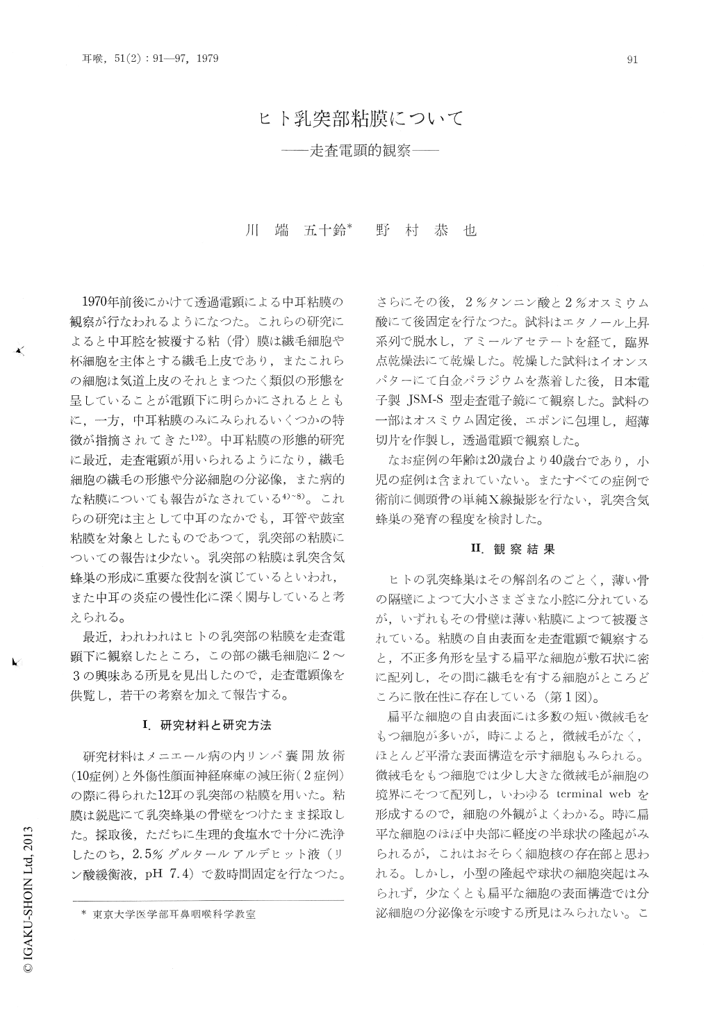
1970年前後にかけて透過電顕による中耳粘膜の観察が行なわれるようになつた。これらの研究によると中耳腔を被覆する粘(骨)膜は繊毛細胞や杯細胞を主体とする繊毛上皮であり,またこれらの細胞は気道上皮のそれとまつたく類似の形態を呈していることが電顕下に明らかにされるとともに,一方,中耳粘膜のみにみられるいくつかの特徴が指摘されてきた1)2)。中耳粘膜の形態的研究に最近,走査電顕が用いられるようになり,繊毛細胞の繊毛の形態や分泌細胞の分泌像,また病的な粘膜についても報告がなされている4)〜8)。これらの研究は主として中耳のなかでも,耳管や鼓室粘膜を対象としたものであつて,乳突部の粘膜についての報告は少ない。乳突部の粘膜は乳突含気蜂巣の形成に重要な役割を演じているといわれ,また中耳の炎症の慢性化に深く関与していると考えられる。
最近,われわれはヒトの乳突部の粘膜を走査電顕下に観察したところ,この部の繊毛細胞に2〜3の興味ある所見を見出したので,走査電顕像を供覧し,若干の考察を加えて報告する。
Mucous membrane of the mastoid cavity obtained during surgery was observed by scanning and transmission electron microscopy. The mucous membrane consists chiefly of flat polygonal cells without cilia (non-ciliated cell), and ciliated cells which are few and scattered among the non-ciliated cells. Distribution of the ciliated cells in this area seems less than 5%. Characteristics of the cilia were described according to the electron microscopic findings. Furthermore we found that the ciliated cells increased in number in the mucous membrane in traumatic facial nerve palsy which was obtained during decompression operation. Those findings are discussed from a viewpoint of the mucociliary system in the middle ear clearance.

Copyright © 1979, Igaku-Shoin Ltd. All rights reserved.


