Japanese
English
- 有料閲覧
- Abstract 文献概要
- 1ページ目 Look Inside
I.はじめに
X線にて下顎骨の下顎角付近に発見される孤立性で円形または楕円形のX線透過像は,1942年Stafne2)が,"Bone cavities situated near theangle of the mandible"という表題で34例の症例を報告して以来,類似の症例報告が相ついでいる。本症は限局性の骨欠損であり,歯芽とは無関係で,無症状で,報告者により"Static bonedcfect"26)29)32)36),"latent bone cyst"7)30)39),"Idiopathic bone cavity"19)34)など種々の名称で呼ばれ,成因についても定説はなく病態もさまざまである。
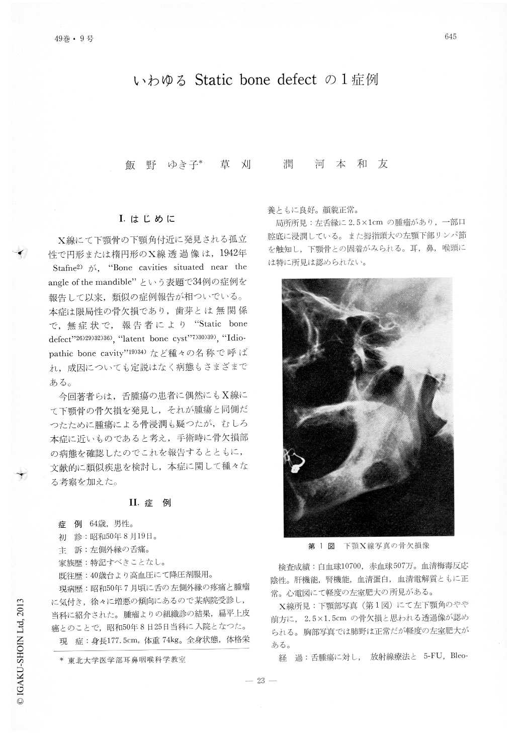
今回著者らは,舌腫瘍の患者に偶然にもX線にて下顎骨の骨欠損を発見し,それが腫瘍と同側だつたために腫瘍による骨浸潤も疑つたが,むしろ本症に近いものであると考え,手術時に骨欠損部の病態を確認したのでこれを報告するとともに,文献的に類似疾患を検討し,本症に関して種々なる考察を加えた。
A case of the so-called "static bone defect" is reported. A man, aged 64 was admitted to the Tohoku University Hospital with the diagnosis of tongue cancer. An X-ray examination on admission revealed an ovoid defect on the body of the mandible near its angle. Sialography showed that this defect was filled with superior lobe of the mandibular salivary gland and this finding was confirmed at surgery.
This disease was initially described and reported by Stafne in 1942 and since then about 90 cases have been reported so far.
This patient happened to have this bony defect on the same side upon which the tongue tumor occurred; bony metastasis of the tumor was initially suspected.

Copyright © 1977, Igaku-Shoin Ltd. All rights reserved.


