Japanese
English
- 有料閲覧
- Abstract 文献概要
- 1ページ目 Look Inside
Ⅰ.緒言
臨床医学における放射性同位元素(以下RIと略す)利用の研究は最近非常に進歩し,癌親和性RIの探索,RI利用による悪性腫瘍の診断が順次進められている。
上顎癌の診断には局所ならびにX線所見,試験切片による病理組織学的検査が行なわれてきた。しかし上顎癌の侵襲範囲はしばしばその洞内にとどまらず骨を破壊して隣接する軟部組織領域ことに眼窩内,翼口蓋窩,頬部筋層などに広汎に波及している場合が多い。このような症例の腫瘍侵襲状態の探知にはX線による特殊撮影(立体,断層撮影,血管造影法など)により側面からその一部をうかがうこともできるが,さらにこれを明らかにする事は単に診断だけではなく治療に際しても重要な意義を持つている。
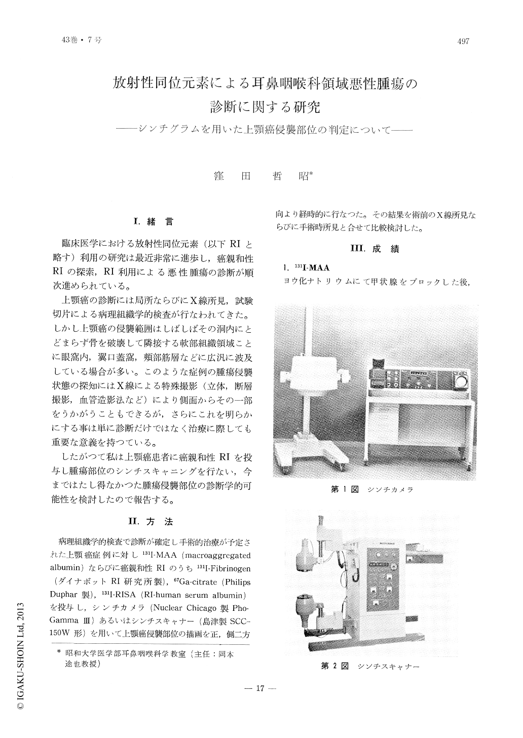
したがつて私は上顎癌患者に癌親和性RIを投与し腫瘍部位のシンチスキャニングを行ない,今まではたし得なかつた腫瘍侵襲部位の診断学的可能性を検討したので報告する。
For the purpose of making diagnosis of cancer of the maxillary region the following studies were made.
With patients suspected of maxillary cancer, a scintigram is taken by means of scintillation camera at intervals of 12, 24, and 48 hours, following intravenous injection of several radioisotopes,I131-RISA, I131-MAA, I131-fibrinogen and Ga67-citrate.
In cases in whom I131-fibrinogen and Ga67-citrate were administered the positive delineation of the growth stood out remarkably well in the scintigram taken 24 hours after the injection of the agents.
The scintigram delineation of the cancer was confirmed at the time of the operation to represent the extent to which the growth actually had occupied.
The RI-contents of the normal and cancerous tissues were measured by means of scintillation counter. Cancer tissues showed a greater accumulation of RI-contents.

Copyright © 1971, Igaku-Shoin Ltd. All rights reserved.


