Japanese
English
- 有料閲覧
- Abstract 文献概要
- 1ページ目 Look Inside
I.はじめに
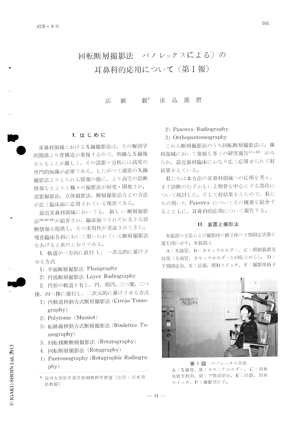
耳鼻科領域におけるX線撮影法は,その解剖学的関係より骨構造が重複するので,明確なX線像をえることが難しく,その読影・分析には高度の専門的知識が必要である。したがつて通常のX線撮影法よりえられる情報の他に,より高度の診断情報をえようと種々の撮影法が研究・開発され,造影撮影法,立体撮影法,断層撮影法などの方法が広く臨床面に応用されている現状である。
最近耳鼻科領域においても,新しい断層撮影法16)18)19)が紹介され,臨床面でそれぞれ大きな診断情報を提供し,その実用性が実証されてきた。現在臨床各科において用いられている断層撮影法をあげると次のとおりである。
1.軌道が一方向に直行し,一次元的に暈けさせる方式
1)平面断層撮影法Planigraphy
2)円孤断層撮影法Layer Radiography
2.円形の軌道を有し,円,楕円,三つ葉,三つ捲,四つ捲に運行し,二次元的に暈けさせる方式
1)円軌道移動方式断層撮影法(Circus Tomography)
2)Polytome(Massiot)
3)転跡線移動方式断層撮影法(Roulettes Tomography)
3.回転横断断層撮影法(Rotatography)
4.回転断層撮影法(Rotagraphy)
1)Pantomography(Rotagraphic Radiography)
2)Panorex Radiography
3)Orthopantomography
Panorex is heavily employed in dentistry but the authors attempted it use in the field of otolaryngology. With panorex it is possible to film the entire dental structures as well as the mandibular joint, the maxillary bone and the sinus, all, on a single plate.
When the target center is slightly raised in the film taking directed at the center of the maxillary bone, it was possible to discern clearly the structures such as the anterior and posterior surface of the maxillary bone, various aspects of the optic fossa surface, the pterygoid process as well as the adjoining pterygopalatine fossa.
In order to be more familiar to the various landmarks panorex of the dry skull was taken in comparison to that of the living one.

Copyright © 1970, Igaku-Shoin Ltd. All rights reserved.


