Japanese
English
- 有料閲覧
- Abstract 文献概要
- 1ページ目 Look Inside
Ⅰ.緒言
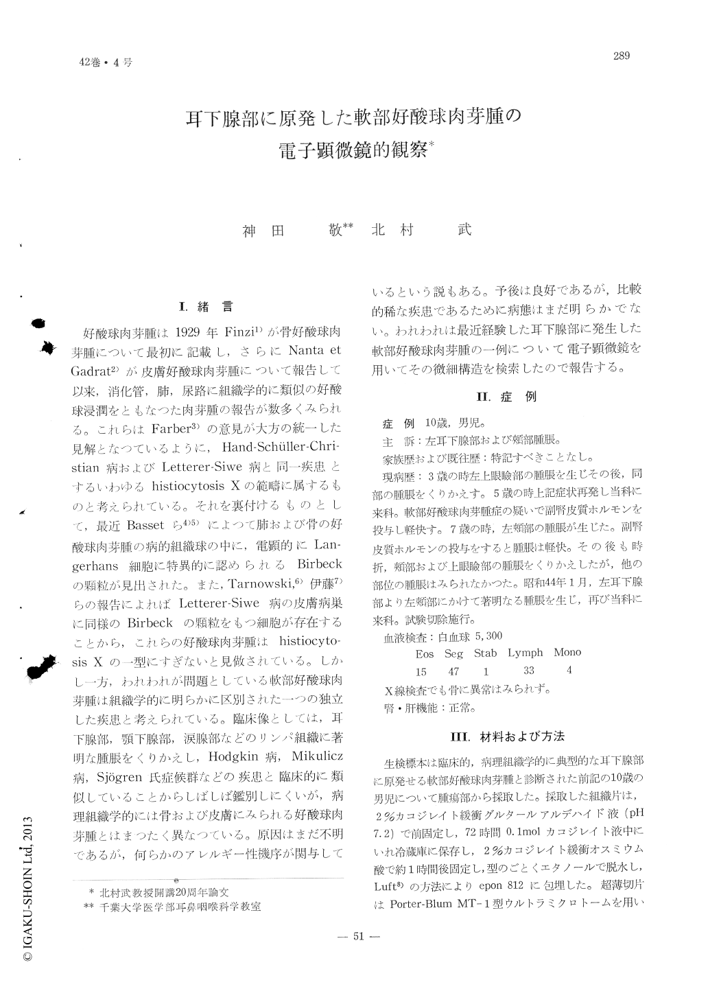
好酸球肉芽腫は1929年Finzi1)が骨好酸球肉芽腫について最初に記載し,さらにNanta et Gadrat2)が皮膚好酸球肉芽腫について報告して以来,消化管,肺,尿路に組織学的に類似の好酸球浸潤をともなつた肉芽腫の報告が数多くみられる。これらはFarber3)の意見が大方の統一した見解となつているように,Hand-Schüller-Christian病およびLetterer-Siwe病と同一疾患とするいわゆるhistiocytosis Xの範疇に属するものと考えられている。それを裏付けるものとして,最近Bassetら4)5)によつて肺および骨の好酸球肉芽腫の病的組織球の中に,電顕的にLangerhans細胞に特異的に認められるBirbeckの顆粒が見出された。また,Tarnowski,6)伊藤7)らの報告によればLetterer-Siwe病の皮膚病巣に同様のBirbeckの顆粒をもつ細胞が存在することから,これらの好酸球肉芽腫はhistiocytosis Xの一型にすぎないと見做されている。しかし一方,われわれが問題としている軟部好酸球肉芽腫は組織学的に明らかに区別された一つの独立した疾患と考えられている。臨床像としては,耳下腺部,顎下腺部,涙腺部などのリンパ組織に著明な腫脹をくりかえし,Hodgkin病,Mikulicz病,Sjögren氏症候群などの疾患と臨床的に類似していることからしばしば鑑別しにくいが,病理組織学的には骨および皮膚にみられる好酸球肉芽腫とはまつたく異なつている。原因はまだ不明であるが,何らかのアレルギー性機序が関与しているという説もある。予後は良好であるが,比較的稀な疾患であるために病態はまだ明らかでない。われわれは最近経験した耳下腺部に発生した軟部好酸球肉芽腫の一例について電子顕微鏡を用いてその微細構造を検索したので報告する。
The eosinophilic granuloma, which involved the parotid gland of a ten-year-male, was studied by light and electron microscopy. The eosinophilic granuloma of the soft tissue is histologically quite different from that of bone and skin This disease is characterized by increase of lymph follicles with germinal center, and eosinophils. Electron microscopical findings showed that lymph follicles consist of lymphoblasts, lymphocytes and eosinophils. Many eosinophils were found aggregatively infiltrating in the vicinity of the lymph follicle. The most of these eosinophils were found to be collapsed and degenerated; the rough endoplasmic reticulum and mitochondria were markedly decreased in number; and, the Golgi apparatus was not prominent The eosinophilic granules were, also, decreased in amount and occasionally showed some abnormal types. Some phagocytotic vesicles, which contained amorphous material, were seen in the cytoplasm of the eosinophils and some Charcot-Leiden crystals were found in the vicinity of the degenerated ones.
However, the eosinophils found in the capilla ry vessels appeared to be normal

Copyright © 1970, Igaku-Shoin Ltd. All rights reserved.


