Japanese
English
- 有料閲覧
- Abstract 文献概要
- 1ページ目 Look Inside
I.はじめに
聴神経鞘腫はしばしば嚢胞を形成するが,腫瘍のほとんどが嚢腫で形成される聴神経鞘腫は現在までに15例ほどしかなく,極めて稀である1-7,9,10,14).今回われわれが経験した症例を含めて,臨床症状,CT,MRI所見,及び病理組織学的所見を検討したので,ここに報告する.
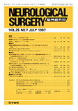
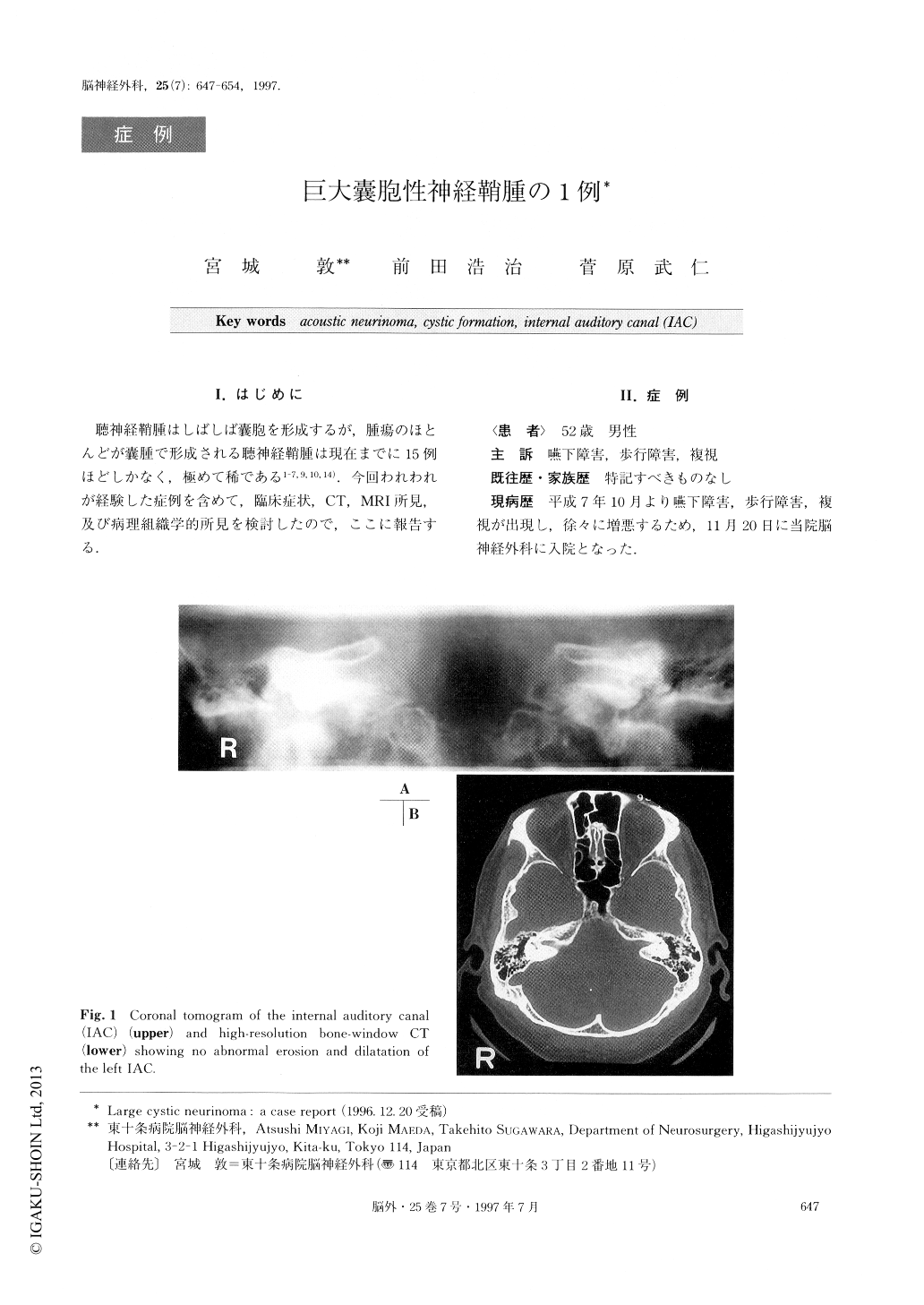
We report a case of large cystic acoustic neurinoma. A 52-year-old male was admitted to hospital with a his-tory of progressive dysphagia, gait disturbance and di-plopia for 2 months. On admission, neurological ex-aminations revealed Bruns' type nystagmus to the left side, hypesthesia in the distribution of the second and third divisions of the left trigeminal nerve, and partial paresis of cranial nerves Ⅸ, Ⅹ, and ⅩⅡ on the left side, and truncal ataxia. A pure-tone threshold audiogram in-dicated the presence of 32dB hearing loss in the left ear. Speech discrimination was 80%. Caloric vestibular responses were absent on the left side. Skull radio-graphs with polytomographs of the internal auditory canal (IAC) were normal. Bony changes in the IAC were not found by high-resolution bone-window com-puted tomography (CT) scan. A plain CT scan re-vealed a large low-attenuated cystic mass in the left cerebellopontine angle (CPA), which was associated with displacement of the fourth ventricle. An enhanced CT scan demonstrated a thin rim-enhancement in the cyst wall. Magnetic resonance imaging (MRI) scans disclosed a large rim-enhanced cystic mass extending superiorly into the tentorial incisura and inferiorly into the foramen magnum. At surgery via a left suboccipitalapproach, a large cystic mass was found at the left CPA arising from the Ⅷth nerve, and compressing the Vth, Ⅵth, Ⅶth and lower cranial nerves. The cyst was filled with a xanthochromic fluid and was firmly attached to the internal auditory meatus (IAM). However no tumor extension into the IAM was con-firmed. The tumor was excised completely. The postop-erative course was uneventful, except for impairment of the Ⅶth and Ⅷth nerves. At 6 months after the first operation, the facial nerve had improved up to grade Ⅲ (Hause-Brackmann stage). Histological examina-tions revealed a typical benign acoustic neurinoma with predominant representation of Antoni B tissues. Thecyst wall contained numerous abnormal sinusoid and telangiectasia-like vessels which showed occasional thromboses. The vessel walls displayed endothelial pro-liferations and were frequently hyalinized. Hemosiderin deposits and hemosiderin-containing phagocytes were also found near these vessels. Myxoid degeneration and necrosis were evident in vast areas. These degenerative changes appeared to be the principal causes of the large cystic formation. 16 cases including our case have been reported. The broad characteristics of the clinical symptoms and radiological findings of these tumors are discussed.
Copyright © 1997, Igaku-Shoin Ltd. All rights reserved.