Japanese
English
- 有料閲覧
- Abstract 文献概要
- 1ページ目 Look Inside
I.はじめに
大後頭孔左前外側部より生じた比較的巨大な髄膜腫に対して経後頭顆到達法により手術を行い,良好な結果を得た.この部位への到達法の術前評価に造影三次元CT scanが有用であったので報告する.
We report a case of foramen magnum meningioma in which case enhanced three-dimensional CT scan was valuable for preoperative evaluation of the surgical ap-proach.
A 53-year-old woman had suffered from stiffness and pain in the left occipital region and numbness of the left side of the face for about 2 years before admission. She had also weakness and numbness of the left side of her body for about 2 months before admission, and dysphagia and pain in the occipital region and in the posterior region of the neck produced by straining for about 1 month before admission. Neurological examina-tion revealed left hemiparesis, and hypalgesia and tac-tile hypesthesia of the left side of the body, including the face.
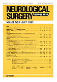
Plain X-P was normal. Enhanced CT scan and gado-linium enhanced MRI revealed a well-enhanced mass attached to the left anterolateral part of the foramen magnum. The left occipital condyle was observed at thelateral side of the attachment part of this mass. Angiography revealed tumor feeders from the menin-geal branches of the left vertebral artery and the left ascending pharyngeal artery.
Enhanced three-dimensional CT scan clearly showed that the tumor was attached to the left anterolateral part of the foramen magnum, that the left occipital con-dyle was at the lateral side of the attachment part of this mass and that the jugular foramen and jugular tubercle were situated superolateral to the attachment part of this mass.
Considering these factors, we decided that removal of the posterior part of the left occipital condyle was necessary, but removal of the left jugular tubercle was not necessary for a good operative view from the left posterior lateral direction.
The tumor was totally removed successfully andgood results were obtained by the transcondylar ap-proach without removal of the jugular tubercle. Histolo-gy of the tumor revealed meningothelial meningioma. In this case, preoperative evaluation with enhanced three-dimensional CT scan was helpful for deciding the surgical approach.
With enhanced three-dimensional CT scan, it is easy to judge whether removal of the posterior part of the occipital condyle and/or the jugular tubercle is neces-sary for a good operative view, and we can get good images revealing the relationships between the tumor and surrounding structues.
Preoperative evaluation with enhanced three-dimensional CT scan is very useful especially in this kind of situation.
Copyright © 1997, Igaku-Shoin Ltd. All rights reserved.