Japanese
English
- 有料閲覧
- Abstract 文献概要
- 1ページ目 Look Inside
I.はじめに
頭蓋底外科手術の手術成績は,最近著しく向上しているが,斜台部脊索腫の手術はいまだにchallengeableな手術であり,全摘が困難なことが多い.脊索腫の予後は,初回の手術で決まるといっても過言ではなく1),いかに全摘がうまくなされるかにかかっている.今回われわれは,神経内視鏡,neuronavigationを併用しながら,摘出しえた症例を経験したので,若干の文献を加えここに報告する.
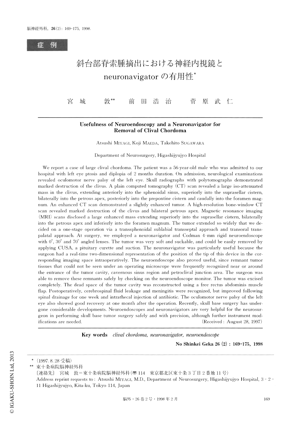
We report a case of large clival chordoma. The patient was a 56-year-old male who was admitted to ourhospital with left eye ptosis and diplopia of 2 months duration. On admission, neurological examinationsrevealed oculomotor nerve palsy of the left eye. Skull radiographs with polytomographs demonstratedmarked destruction of the clivus. A plain computed tomography (CT) scan revealed a large iso-attenuatedmass in the clivus, extending anteriorly into the sphenoidal sinus, superiorly into the suprasellar cistern,bilaterally into the petrous apex, posteriorly into the prepontine cistern and caudally into the foramen mag-num. An enhanced CT scan demonstrated a slightly enhanced tumor. A high-resolution bone-window CTscan revealed marked destruction of the clivus and bilateral petrous apex. Magnetic resonance imaging(MRI) scans disclosed a large enhanced mass extending superiorly into the suprasellar cistern, bilaterallyinto the petrous apex and inferiorly into the foramen magnum. The tumor extended so widely that we de-cided on a one-stage operation via a transsphenoidal sublabial transseptal approach and transoral trans-palatal approach. At surgery, we employed a neuronavigator and Codman 4-mm rigid neuroendoscope with 0°, 30° and 70° angled lenses. The tumor was very soft and suckable, and could be easily removed byapplying CUSA, a pituitary curette and suction. The neuronavigator was particularly useful because thesurgeon had a real-time two-dimensional representation of the position of the tip of this device in the cor-responding imaging space intraoperatively. The neuroendoscope also proved useful, since remnant tumortissues that could not be seen under an operating microscope were frequently recognized near or aroundthe entrance of the tumor cavity, cavernous sinus region and petroclival junction area. The surgeon wasable to remove these remnants safely by checking on the neuroendoscope monitor. The tumor was excisedcompletely. The dead space of the tumor cavity was reconstructed using a free rectus a bdominis muscleflap. Postoperatively, cerebrospinal fluid leakage and meningitis were recognized, but improved followingspinal drainage for one week and intrathecal injection of antibiotic. The oculomotor nerve palsy of the lefteye also showed good recovery at one month after the operation. Recently, skull base surgery has under-gone considerable developments. Neuroendoscopes and neuronavigators are very helpful for the neurosur-geon in performing skull base tumor surgery safely and with precision, although further instrument mod-ifications are needed.

Copyright © 1998, Igaku-Shoin Ltd. All rights reserved.


