Japanese
English
- 有料閲覧
- Abstract 文献概要
- 1ページ目 Look Inside
I.はじめに
心房内粘液腫は脳梗塞や脳動脈瘤を合併し,様々な神経症状をきたすことがあるが,心房内粘液腫に合併した脳動脈瘤について経時的変化を追跡した報告は極めて少ない.今回われわれは心房内粘液腫により発生した脳動脈の症例に対し,手術を施行し,術後もintravenous digital subtraction angiography(IVDSA)にて経時的変化を追跡しえた症例を経験したので報告する.
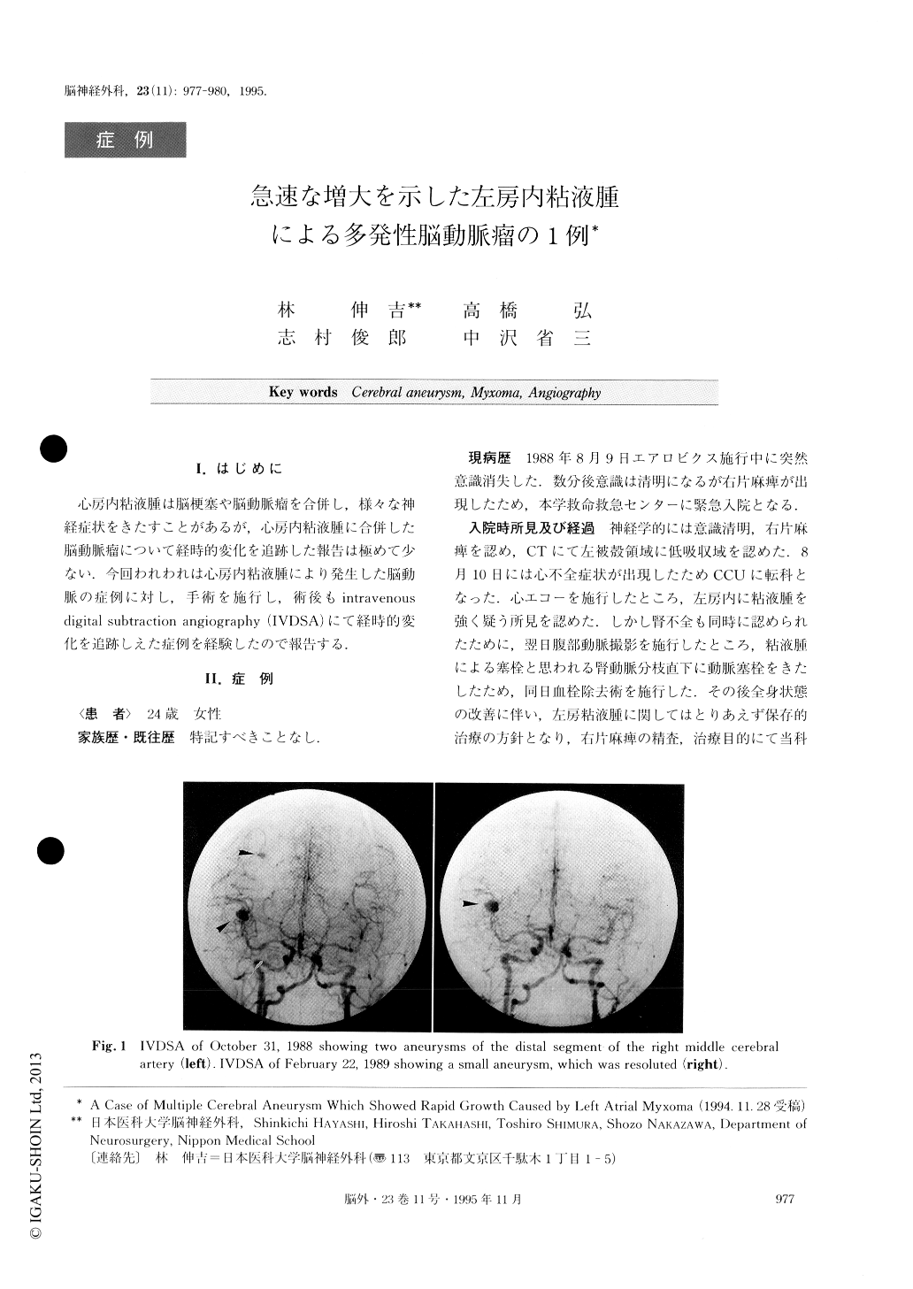
A 24-year-old woman was admitted complaining of right hemiparesis and episodes of syncope. Computed tomography demonstrated a low density area in the left putaminal region. Intravenous digital subtraction angiography (IVDSA) showed two aneurysms in the distal segment of the right middle cerebral artery. Cere-bral emboli from a cardiac source was suspected, and cardioechography was performed. Myxoma was located in the left atrium. The patient was transferred to a car-dio surgical unit, and the myxoma was successfully re-moved. After removal by operation of the cardiac tumor, follow-up third IVDSA was performed. One aneurysm of the distal segment of the right mid-dle cerebral artery had grown larger. On the other hand, the other aneurysm had dissappeared. Clipping of the enlarged aneurysm was performed. After the clip-ping operation of the enlarged aneurysm, a follow-up 4th IVDSA was performed. A new aneurysm of the proximal segment of the left cerebral artery was observed. A follow-up 5th IVDSA was performed, re-vealing that the new aneurysm was enlarging. No op-eration was performed, because the aneurysm was the fusiform type.
At present, the patient is complaining of slight right hemiparesis and has returned to her job. Here we re-ported a case of cerebral aneurysm caused by left atrial myxoma.

Copyright © 1995, Igaku-Shoin Ltd. All rights reserved.


