Japanese
English
- 有料閲覧
- Abstract 文献概要
- 1ページ目 Look Inside
I.はじめに
髄液鼻漏の多くは外傷性で頭部外傷に起因するものが約80%,前頭蓋底腫瘍の手術や耳鼻科手術に続発する術後髄液鼻漏が約16%である.これに対し,非外傷性髄液鼻漏はまれであり,3-4%をしめるにすぎない8).今回,われわれは水頭症により中頭蓋底に瘻孔を形成したきわめてまれな非外傷性髄液鼻漏の1例を経験したので,その病因を中心に若干の文献的考察を加えて報告する.
We report an unusual case of non-traumatic cerebro-spinal fluid rhinorrhea associated with aqueductal steno-sis and hydrocephalus.
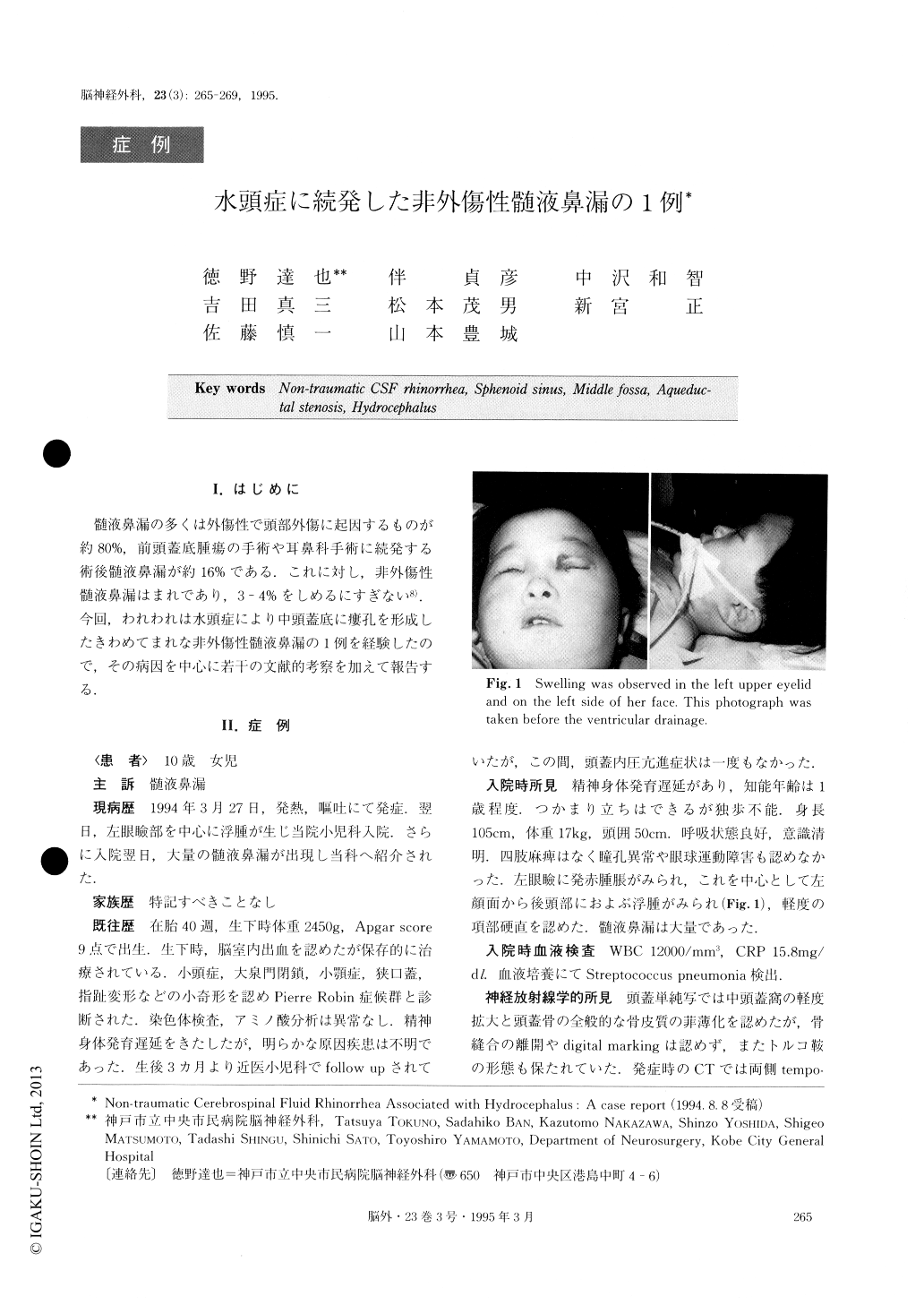
The patient was a 10-year-old girl who suddenly de-veloped massive CSF rhinorrhea following severe ede-ma of the left side of her face. CT scan showed marked dilatation of the lateral and third ventricles and en-larged sphenoid sinus of water density, extending to the lateral wall of the left orbit and to the left ptery-goid fossa. Immediately after the onset of CSF rhinor-rhea, ventricular drainage was performed, but the rhi-norrhea persisted. Ventriculography revealed predomi-nant flow of the contrast medium into the left temporal horn and abnormal collection in the sphenoid sinus. Coronal CT scan did not show any focal bony defect, but a thin layer was seen in the base of the left middle fossa. Exploration of the skull base in the left middle fossa was performed through a left frontotemporal cra-niotomy. An irregular bony defect measuring 7 × 12mm was then found in the anterolateral floor of the middle fossa and the dura was also perforated there. Brain tis-sue including the temporal horn protruded through the bony defect into the sphenoid sinus. After excision of the herniated brain tissue, repair was accomplished by packing muscle into the bony defect and covering the dural defect with fat reinforced by coating with fibrin glue. Postoperatively, the CSF rhinorrhea has stopped and the edema of her face has disappeared.
We discuss the etiology of this unusual spontaneous CSF leakage through the middle fossa and the abnor-mally enlarged sphenoid sinus.

Copyright © 1995, Igaku-Shoin Ltd. All rights reserved.


