Japanese
English
- 有料閲覧
- Abstract 文献概要
- 1ページ目 Look Inside
I.はじめに
小児において軽微な頭部外傷後,大脳基底核または深部白質の中大脳動脈穿通枝領域に梗塞巣の出現をきたし得ることが知られているが4,6,9),その機序については推論の域を出ていない.今回われわれは軽微な頭部外傷後に続発した小児脳梗塞の1例を経験したが,神経放射線学的に興味ある所見を呈したので報告するとともに脳梗塞発症機序について考察した.
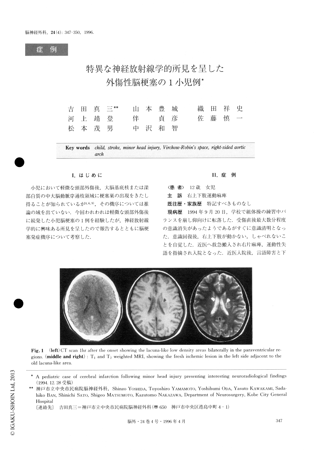
We reported a case of juvenile cerebral infarction fol-lowing minor head injury. The patient, a 12-year-old girl, developed right hemiparesis and aphasia almost immediately after having fallen from about 1 meter height during the exercise class at school. CT and MRI study showed lacunar lesions bilaterally and almost symmetrically in the paraventricular deep white matter on both sides. A new stroke area, responsible for the symptoms, was recognized about 24 hours later on CT scan just next to the lacuna of the left side. Although angiography revealed a rare anomaly of the right side aortic arch, associated with subclavian steal phe-nomenon, presumably of congenital in nature, no abnor-mality was found in the intracranial vessels. She made a rapid recovery during her hospital stay and showed no more than a slight motor weakness in her right up-per extremity on discharge. The literature was re-viewed on the embryology of the aortic arch and brachiocephalic arteries. We speculate that the lacunar lesions found bilaterally are dilated large normal Vir-chow-Robin space, and the pathogenesis of the stroke in this patient was discussed.

Copyright © 1996, Igaku-Shoin Ltd. All rights reserved.


