Japanese
English
- 有料閲覧
- Abstract 文献概要
- 1ページ目 Look Inside
I.はじめに
平滑筋肉腫は平滑筋組織から生ずる稀な悪性腫瘍であり,消化器,子宮,後腹膜,皮下などに好発する.転移は肺と肝臓に多く,頭蓋内への転移は稀である.また骨への転移も稀で,とくに頭蓋骨への転移については組織学的に確認されている報告はKomataら8)の頭蓋冠へ転移した1例のみであり,蝶形骨への転移例は見当たらない.われわれは眼球突出と側頭部腫瘤を主訴として来院した眼窩内伸展を伴う側背部皮下平滑筋肉腫の蝶形骨転移例を経験し,極めて稀と考えられたので若干の文献的考察を加えて報告する.
A rare subcutaneous leiomyosarcoma metastatizing to the sphenoid bone and presenting exophthalmos is reported. A 56-year-old female presented with protru-sion of the right eye and a slowly growing lump on the right temporal region. Six years previously, she had undergone removal of a subcutaneous mass in the back, which was histologically diagnosed in another hospital as leiomyosarcoma. She had undergone four other op-erations, including removal of local recurrences and a right renal metastasis.
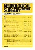
On admission, physical examination showed no neurological deficits. Craniogram revealed an osteolytic lesion without marginal sclerosis in the right sphenoid bone. CT showed an inhomogeneously enhanced mass with irregular expansion of the diploic space, which was partly invading the right orbit. MRI demonstrated an extradural mass in the right sphenoid region, which was slightly low-intense in T1-weighted image, high-intense in T2-weighted image, and inhomogeneously en-hanced by Gd-DTPA. Right external carotid angiogram showed a highly vascular stain fed by meningeal arte-ries. Radionuclide bone scintigram showed multiple high-uptake areas in the left femoral head, the ribs, and the sphenoid bone.
Preoperative embolization of the tumor vessels fed by the external carotid artery was performed. Follow-ing this procedure, the tumor stain disappeared com-pletely. The tumor was totally excised with minimal bleeding through an orbitozygomatic approach. The tumor was loosely adherent to the dura and periorbit. The bone defect was covered with a methylmethacry-late resin plate. The histological examination demon-strated fascicular arrangement of the spindle shaped cells with mitotic figures. Immunohistochemical studies showed that most tumor cells were positive for actin and myosin, but negative for desmin. Accordingly the tumor was diagnosed as leiomyosarcoma. The patient followed a satisfactory postoperative course without neurological deficits. However, she died of general dis-semination of the tumor in another hospital 10 months after the craniotomy.
Leiomyosarcomas arise most commonly in the gas-trointestinal and female genital tract; other viscera, major arteries, veins, and the extremities are less fre-quent sites of origin. Neither chemotherapy nor radiotherapy is apparently effective. Leiomyosarcomas aggressively invade surrounding tissues and dissemi-nate hematogenously, most frequently to the lung. The metastasis of leiomyosarcoma to the central nervous system is a rare event. Only thirteen cases of pathologi-cally confirmed cerebral metastasis of leiomyosarcoma are reported in the literature. Furthermore, only one case of skull metastasis of leiomyosarcoma is reported. With this report, a new case is added to the exceptional observations about skull metastasis of leiomyosarcoma. We think that such patients in good physical condition should be treated surgically to maintain a good quality of life, even in the presence of multiple metastases.
Copyright © 1995, Igaku-Shoin Ltd. All rights reserved.