Japanese
English
- 有料閲覧
- Abstract 文献概要
- 1ページ目 Look Inside
I.はじめに
前頭骨から蝶形骨にかけて発生するfibrous dysplasia(以下FD)は顔面の変形や眼球の偏位のみならず脳神経を巻き込み視力,視野障害を中心とした多彩な眼症状を引き起すため積極的な治療を必要とする疾患である.本疾患の治療時期に関しては良性疾患であり思春期以降に発育の自然停止をみる例が多いことや,すでに生じた視力障害に視神経管開放術が必ずしも有効でないことなどから症状が進行するまで手術期を遅らす傾向がみられた.しかし最近では顕微鏡手術の進歩に伴い慢性的な視神経の圧迫による視力障害であっても視神経管開放術による視力改善の効果が望めるようになり本疾患の手術時期の検討が必要であると思われる.今回われわれは視力障害のない視神経管狭窄を伴うfronto-orbital FDの1例に対し頭蓋・眼窩形成術と同時に視神経管開放術を行い良好な結果を得ることができたので報告する.
A surgical case of monostotic fibrous dysplasia of the left frontal and sphenoidal bone in a 14-year-old girl is described.
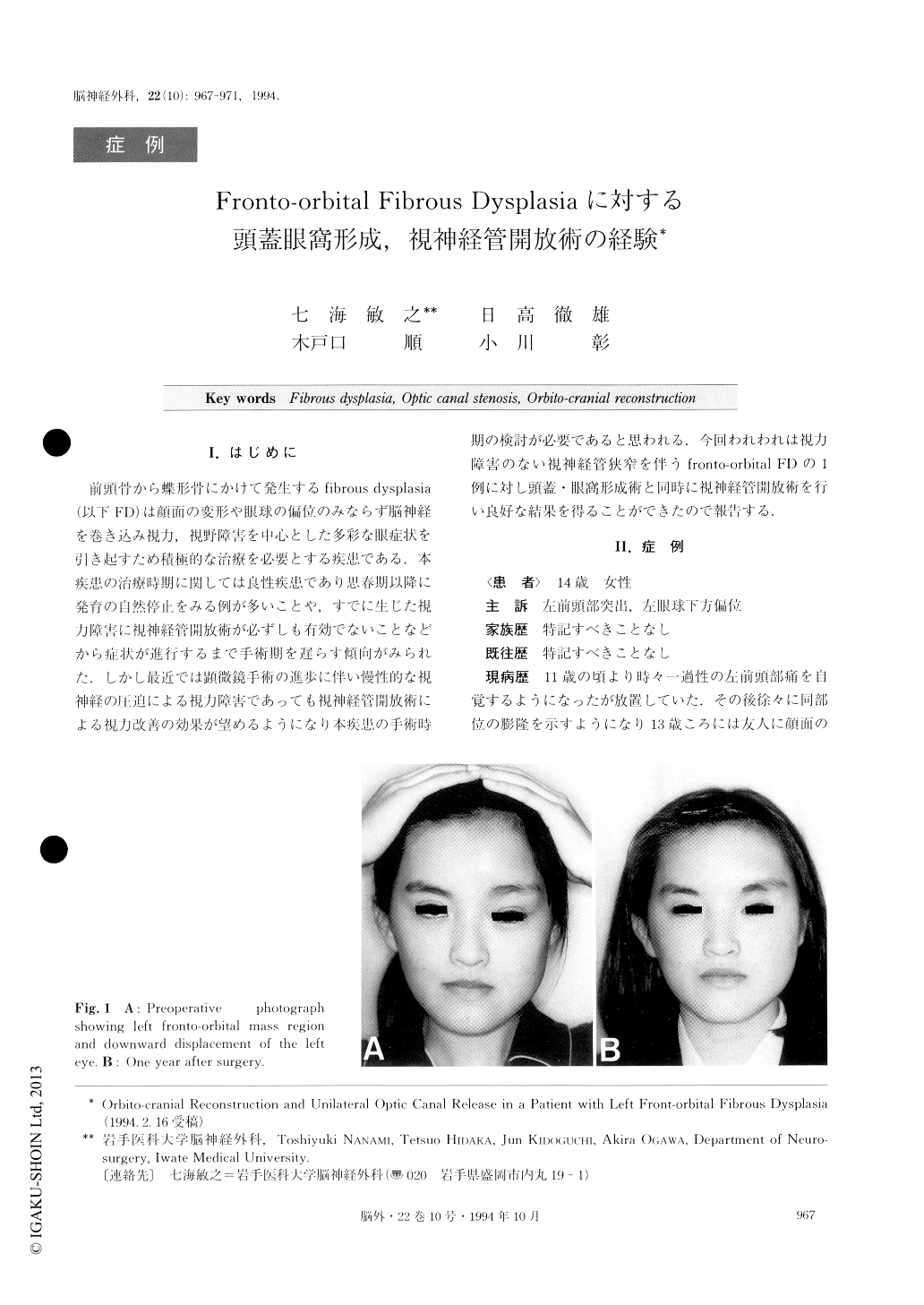
This girl was admitted to our hospital in March, 1992, with a chief complaint of facial deformity and asymmetry due to a painless and progressive bony bulging over the left fronto-orbital region. But she de-nied any symptoms such as proptosis, diplopia, optic atrophy and visual loss. Other data found on neurolo-gical examination and laboratory tests were normal. In addition, she had no history of skin lesions, precocious puberty or other endocrine abnormalities.
Plain craniogram showed remarkable thickening of the left frontal bone and of the anterior cranial fossa of the sphenoidal bone with irregular stenosis of the left optic canal. CT scan showed the diffuse enlargement of the affected bone and involvement of the paranasal sinuses. Angiography revealed no positive findings. On December 10, 1992, orbito-cranial reconstruction and unilateral optic canal release were performed using an extradural approach through a left fronto-temporal cra-nietomy.
Histological fingings confirmed the lesion to be typic-al fibrous dysplasia. She recovered completly one month after the operation, but she suffered transient blurred vision, diplopia and left ptosis. Most of the decreased vision caused by fibrous dys-plasia cannot be reversed after surgical treatment. So, if optic canal stenosis is evident, even when visual loss is not clear, release of the optic canal stenosis should be done as early as possible in association with experi-enced neurosurgeons and with meticulous dissection.

Copyright © 1994, Igaku-Shoin Ltd. All rights reserved.


