Japanese
English
- 有料閲覧
- Abstract 文献概要
- 1ページ目 Look Inside
I.はじめに
頭蓋内髄膜腫はクモ膜細胞由来の腫瘍で,通常硬膜もしくは脳室脈絡叢に付着して存在する.しかし稀にはこのような付着をもたない例も存在することが知られている4).
最近われわれは髄膜腫としては非典型的な画像所見を呈し手術所見より硬膜付着をもたず,また病理組織学的にも興味ある所見を呈した後頭蓋窩髄膜腫の1例を経験したので若干の文献的考察を加え報告する.
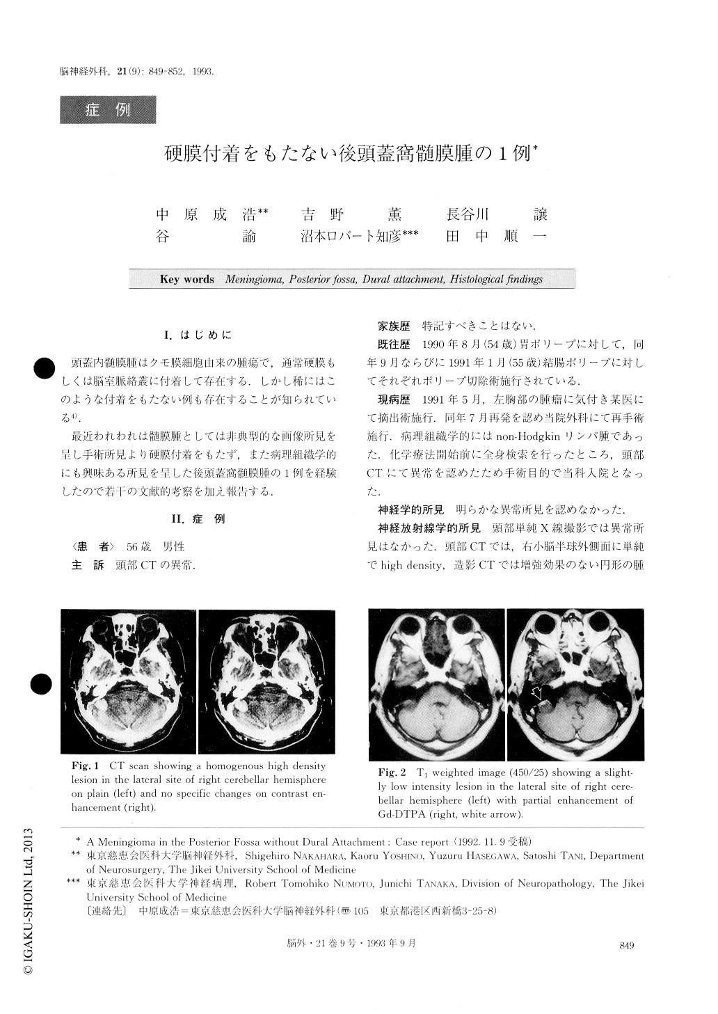
An extremely rare case of a meningioma in the pos-terior fossa without dural attachment has been reported. The patient was a 56-year-old male whose chief man-ifestation was the abnormality of his CT scan. His past history included gastric and colonic polyp when he was 54, 55 years old, and non-Hodgkin's lymphoma before hospitalization in our department. CT scan showed a small round non-enhancing lesion located at the lateral site of the right cerebellar cortex. T1 weighted image of MRI showed a homogenous low intensity lesion with partial enhancing with Gd-DTPA.Proton image showed a remarkable low intensity lesion which showed an extramedullary mass. Right retromastoid craniec-tomy was performed. The mass was an extramedullary tumor which had no relation with the cerebellar cortex and dura matter. The arachnoid membrane around the tumor was intact. The tumor was totally resectecl and the patient had no neurological deficits.
Histopathologically, the tumor was delineated into laminar structures by collagen fiber. Tumor cells were spindle in shape and made a whorling formation. There was no psammoma body and it had a hyperchromatic nuclei without mitotic features. Electron microscopic stu-dies revealed no typical interdigitation but irregularity of the cell membrane. Abundant collagen fibers were in contact with basement membrane of the tumor. Accord-ing to these findings, we diagnosed fibroblastic menin-gioma with atypical forms.
Meningiomas without dural attachment are rare in adults, especially extremely rare of the posterior fossa. There are only 23 previous reports of “meningioma of the posterior fossa without dural attachment”. Cantore divided these meningiomas into three groups (IV ventri-cle, inferior tela choroidea and cisterna magna). Our pre-sent case had no attachment to the dura mater and chor-oid plexus of IV ventricle.
We consider that this meningioma occurred in the col-lagen fiber mass or arose from the ectopic dural sinus with abundant collagen fibers which revealed no dural attachment and showed an atypical MR image of meningioma.
We conclude that our case falls under another cate-gory, that of meningioma of the posterior fossa without dural attachment.

Copyright © 1993, Igaku-Shoin Ltd. All rights reserved.


