Japanese
English
- 有料閲覧
- Abstract 文献概要
- 1ページ目 Look Inside
I.はじめに
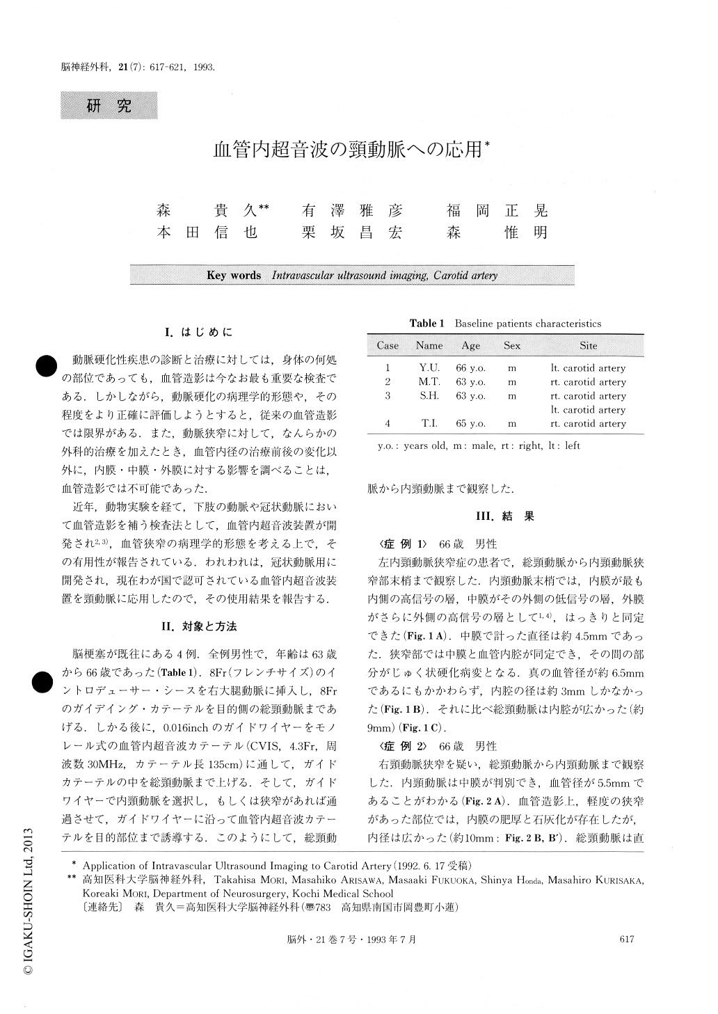
動脈硬化性疾患の診断と治療に対しては,身体の何処の部位であっても,血管造影は今なお最も重要な検査である.しかしながら,動脈硬化の病理学的形態や,その程度をより正確に評価しようとすると,従来の血管造影では限界がある.また,動脈狭窄に対して,なんらかの外科的治療を加えたとき,血管内径の治療前後の変化以外に,内膜・中膜・外膜に対する影響を調べることは,血管造影では不可能であった.
近年,動物実験を経て,下肢の動脈や冠状動脈において血管造影を補う検査法として,血管内超音波装置が開発され2,3),血管狭窄の病理学的形態を考える上で,その有用性が報告されている.われわれは,冠状動脈用に開発され,現在わが国で認可されている血管内超音波装置を頸動脈に応用したので,その使用結果を報告する.
Although conventional angiography is utilized to assess the extent and severity of carotid artery disease, it yields only a silhouette of the vessel lumen. Intravascular ultrasound imaging (IUI), which has been developed for imaging the coronary artery, can supplement angiogra-phy by providing a tomographic perspective of the vessel wall structure. Therefore, we applied IUI (4.3F, 30MHz) to the carotid artery to evaluate the extent of arteriosc-lerosis, and we were successful in imaging the perspec-tive of the carotid artery.
Copyright © 1993, Igaku-Shoin Ltd. All rights reserved.