Japanese
English
症例
視神経交叉部Actinomycosisの1例
Actinomycotic Granuloma of the Optic Chiasma: A case report
豊田 収
1
,
野尻 健
1
,
中島 英雄
1
Osamu TOYODA
1
,
Ken NOJIRI
1
,
Hideo NAKAJIMA
1
1中央群馬脳神経外科病院脳神経外科
1Department of Neurosurgery, Central Gunma Neurosurgical Hospital
キーワード:
Cerebral actinomycosis
,
Optic nerve and chiasma
,
Granuloma
Keyword:
Cerebral actinomycosis
,
Optic nerve and chiasma
,
Granuloma
pp.623-627
発行日 1993年7月10日
Published Date 1993/7/10
DOI https://doi.org/10.11477/mf.1436900673
- 有料閲覧
- Abstract 文献概要
- 1ページ目 Look Inside
I.はじめに
Actinomycosisは化学療法の発達につれ減少の一途をたどっていると言われ,更に中枢神経系のactinomy—cosisは非常に稀な疾患である.今度感染症の徴候に乏しく,術前の諸検査で脳腫瘍と思われたが,術後病理学的に初めて視交叉部と視神経に限局したactinomycosisと判明し,抗生剤の長期投与にて治癒できた1例を経験した.その診断,治療の問題点について考察した.
A case of actinomycotic granuloma of the optic chias-ma and the optic nerve is reported.
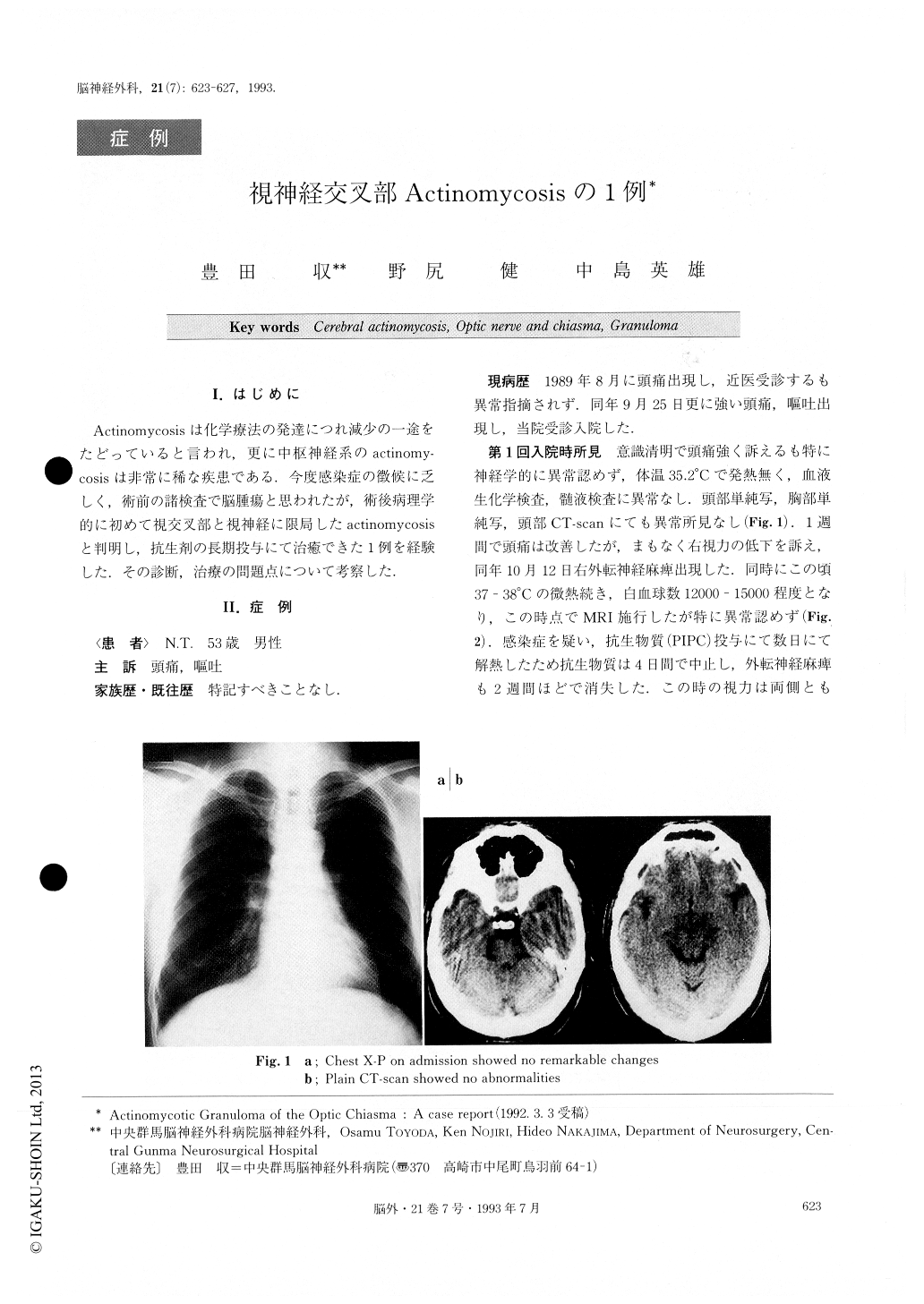
A 53-year-old man was admitted to our hospital with headache and vomiting on September 25, 1989. Generalphysical and neurological examination on admission re-vealed no remarkable findings. CT-scan demonstrated almost normal pictures. On the 17th hospital day, his temperature was 38℃ and white blood cell (WBC) count was 12000 cumm. And he presented right abducens pal-sy. MRI demonstrated no abnormal findings then.

Copyright © 1993, Igaku-Shoin Ltd. All rights reserved.


