Japanese
English
- 有料閲覧
- Abstract 文献概要
- 1ページ目 Look Inside
I.はじめに
前篩骨動脈をfeederとする頭蓋内AVMは,1963年にLepoireらによってはじめて報告され5),現在まで文献上27例の報告がなされている.これらの大多数は,nidusが硬膜に存在する硬膜動静脈奇形(以下duralAVM)である.一方,Teradaらは前篩骨動脈がfeederであるものの,nidusが脳実質内に存在する非定型例を1984年に報告し9),以来3例の報告1,8,10)がなされている.今回われわれも,前篩骨動脈がfeederとなりnidusが皮質に存在した頭蓋内動静脈奇形を経験し極めて稀な症例と考えられたので,文献的考察を加えて報告する.
Only 27 cases of dural AVM fed by the anterior ethmoid artery have been reported in the literature. Their nidi were usually in the dura mater. In our case, the nidus was located in the brain parenchyma, although its feeder was the dural artery.
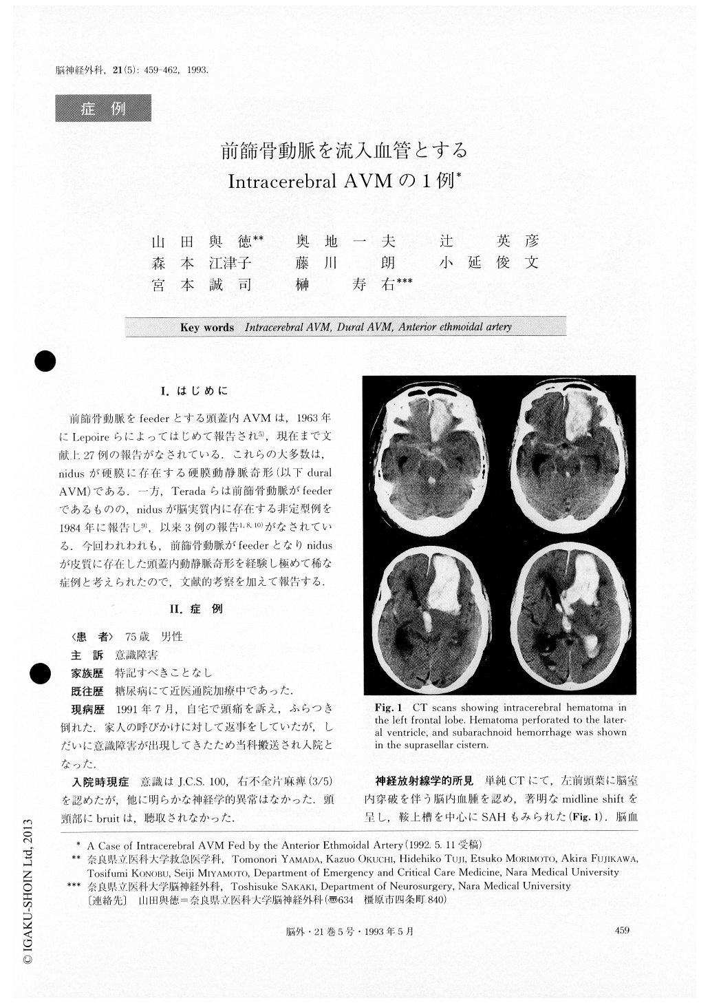
A 75-year-old man was admitted to our department be-cause of disturbed consciousness. CT scan showed in-tracerebral hemorrhage in the left frontal region with ventricular perforation, and subarachnoid hemorrhage in the suprasellar cistern. Left carotid angiography revealed an AVM in the anterior cranial fossa, fed by the anterior ethmoidal artery and drained by a cortical vein, which was dilated with some vascular sacs.
A left frontal craniotomy was performed. The subcor-tical hematoma was removed, and then, after retracting the frontal lobe, two feeders penetrating the dura mater were identified and clipped. The nick's of the AVM with aneurysmal vascular dilatation could be seen on the cor-tical surface. It was coagulated and removed enbloc. His-tologically, the malformation consisted of thickened di-lated veins and distorted small arteries. Postoperative angiography revealed no vascular anomaly.
The patient was discharged with mild aphasia and mild right hemiparesis.

Copyright © 1993, Igaku-Shoin Ltd. All rights reserved.


