Japanese
English
- 有料閲覧
- Abstract 文献概要
- 1ページ目 Look Inside
I.はじめに
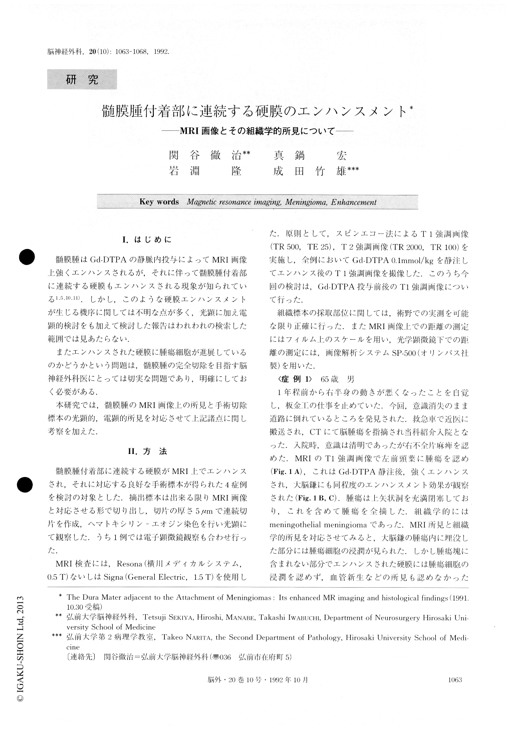
髄膜腫はGd-DTPAの静脈内投与によってMRI画像上強くエンハンスされるが,それに伴って髄膜腫付着部に連続する硬膜もエンハンスされる現象が知られている1,5,10,11).しかし,このような硬膜エンハンスメントが生じる機序に関しては不明な点が多く,光顕に加え電顕的検討をも加えて検討した報告はわれわれの検索した範囲では見あたらない.
またエンハンスされた硬膜に腫瘍細胞が進展しているのかどうかという問題は,髄膜腫の完全切除を目指す脳神経外科医にとっては切実な問題であり,明確にしておく必要がある.
本研究では,髄膜腫のMRI画像上の所見と手術切除標本の光顕的,電顕的所見を対応させて上記諸点に関し考察を加えた.
The dura mater adjacent to the attachment of mening-iomas was enhanced on MR imaging with intravenous Gd-DTPA infusion. It was examined histologically in four patients with intracranial globoid meningiomas. His-tological examination revealed that there was no tumor cell invasion within the dura mater enhanced on MR im-aging, except at the point of their attachment. A layer of tumor cells was occasionally observed on the surface of the dura mater, but this was limited to within 5 mm of the tumor margin.
Our electronmicroscopic observation indicated that en-hancement of the dura mater adjacent to the attachment of meningiomas was caused by increased vascular per-meability of the dural vessels and extended extravascular space.

Copyright © 1992, Igaku-Shoin Ltd. All rights reserved.


