Japanese
English
- 有料閲覧
- Abstract 文献概要
- 1ページ目 Look Inside
I.はじめに
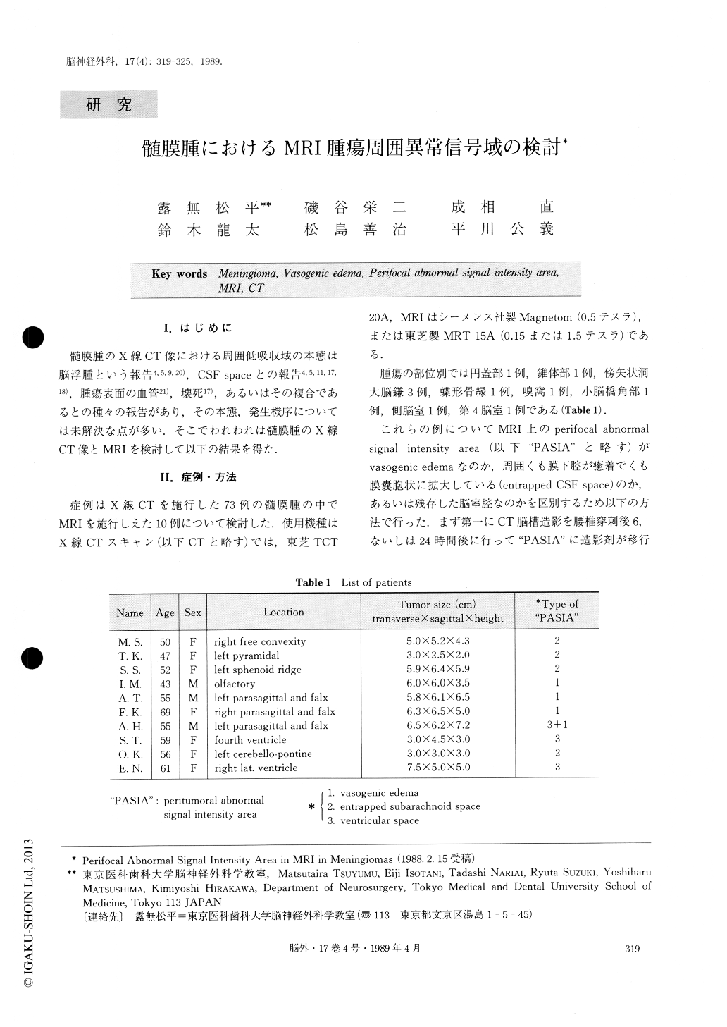
髄膜腫のX線CT像における周囲低吸収域の本態は脳浮腫という報告 4,5,9,20),CSF spaceとの報告4,5,11,17,18),腫瘍表面の血管21),壊死17),あるいはその複合であるとの種々の報告があり,その本態,発生機序については未解決な点が多い.そこでわれわれは髄膜腫のX線CT像とMRIを検討して以下の結果を得た.
To investigate the perifocal abnormal signal intensity area in MRI in meningiomas, we have analysed MRI in 10 cases among 73 meningiomas which were diagnosed by X-ray CT and verified by operation and pathology.
The MRI scanners used in this study were Siemens Magnetom and Toshiba MRT 15A. Ten meningiomas diagnosed by MRI were as follows ; one free convex-ity, one pyramidal, three falx and parasagittal, one sphenoid ridge, one olfactory groove, one cerebello-pontine angle, two ventricular meningiomas. Perifocal abnormal signal intensity area was diagnosed as vasogenic edema in 4 cases.

Copyright © 1989, Igaku-Shoin Ltd. All rights reserved.


