Japanese
English
- 有料閲覧
PPV購入案内
1,320円 (1,200円+税10%) こちらは、715ページから721ページの文献です。購入には医書.jp本会員登録が必要です。
会員登録完了後に改めて上記「購入サイトへ」を選択してください。
または「購入サイトへ」選択後に「購入ログイン」、「新規会員登録」の順に進んでください。
会員登録について詳しくはをご確認ください。
- Abstract 文献概要
- 1ページ目 Look Inside
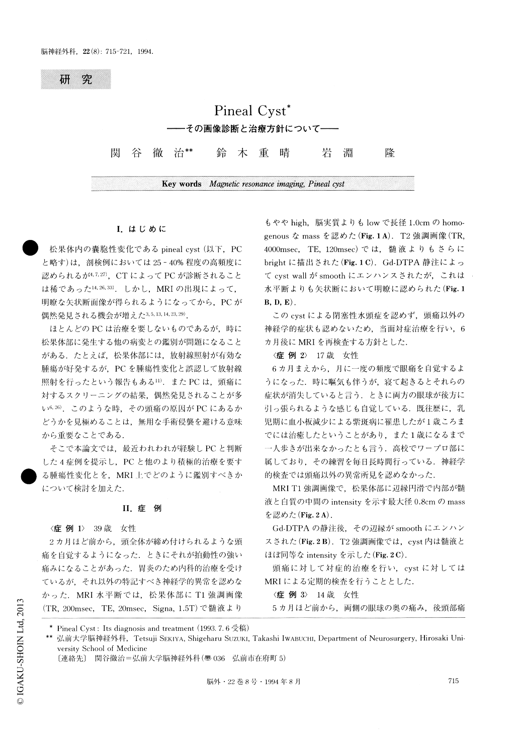
I.はじめに
松果体内の嚢胞性変化であるpineal cyst(以下,PCと略す)は,剖検例においては25-40%程度の高頻度に認められるが4,7,27),CTによってPCが診断されることは稀であった14,26,33).しかし,MRIの出現によって,明瞭な矢状断面像が得られるようになってから,PCが偶然発見される機会が増えた3,5,13,14,23,29).
ほとんどのPCは治療を要しないものであるが,時に松果体部に発生する他の病変との鑑別が問題になることがある.たとえば,松果体部には,放射線照射が有効な腫瘍が好発するが,PCを腫瘍性変化と誤認して放射線照射を行ったという報告もある11).またPCは,頭痛に対するスクリーニングの結果,偶然発見されることが多い6,26).このような時,その頭痛の原因がPCにあるかどうかを見極めることは,無用な手術侵襲を避ける意味から重要なことである.
Four cases of pineal cyst were presented and their fea-tures on magnetic resonance (MR) imaging were dis-cussed while reviewing the literature relevant to this clinical entity.
Pineal cyst is a normal developmental variant and in most of the cases its diagnosis is not difficult, but in a limited number of cases, pineal cyst needs to be differ-entiated from cystic tumors which have developed in the region of the pineal gland, such as pineocytoma or astrocytoma. Most pineal cysts are found incidentally during MR screening studies and no surgical interven-tions are needed. Pineal cysts should be considered for surgery only when the diameter of the cyst is more than 1.0cm, or when obstructive hydrocephalus is veri-fied on MRI or CT, and/or when definitive neurologic-al signs such as Parinaud's syndrome are present. It was also stressed that embryological considera-tions are indispensable for evaluating pineal cysts on MR images.
Copyright © 1994, Igaku-Shoin Ltd. All rights reserved.