Japanese
English
- 有料閲覧
- Abstract 文献概要
- 1ページ目 Look Inside
I.はじめに
頭蓋内解離性動脈瘤は,従来比較的稀とされてきたが近年その報告は増加し,以前考えられていた以上に多い疾患と言える.特に,椎骨・脳底動脈領域では,クモ膜下出血で発症しやすいことが指摘されている.
今回,われわれはクモ膜下出血で発症した解離性椎骨動脈瘤で,急性期の脳血管撮影では,解離性動脈瘤が見出しえず,再度の脳血管撮影で初めて診断が出来た稀な症例を経験した.本症例の詳細を報告し,あわせて文献的考察を行う.
Dissecting aneurysm of the intracranial arteries is a well known clinical entity, and its angiographic findings are also well recognized. We encountered a case with dissecting aneurysm of the vertebral artery presented with subarachnoid hemorrhage (SAH). The initial angiography was normal but repeated angiography de-monstrated a dissecting aneurysm. This case is reported here, and the relevant literature is reviewed.
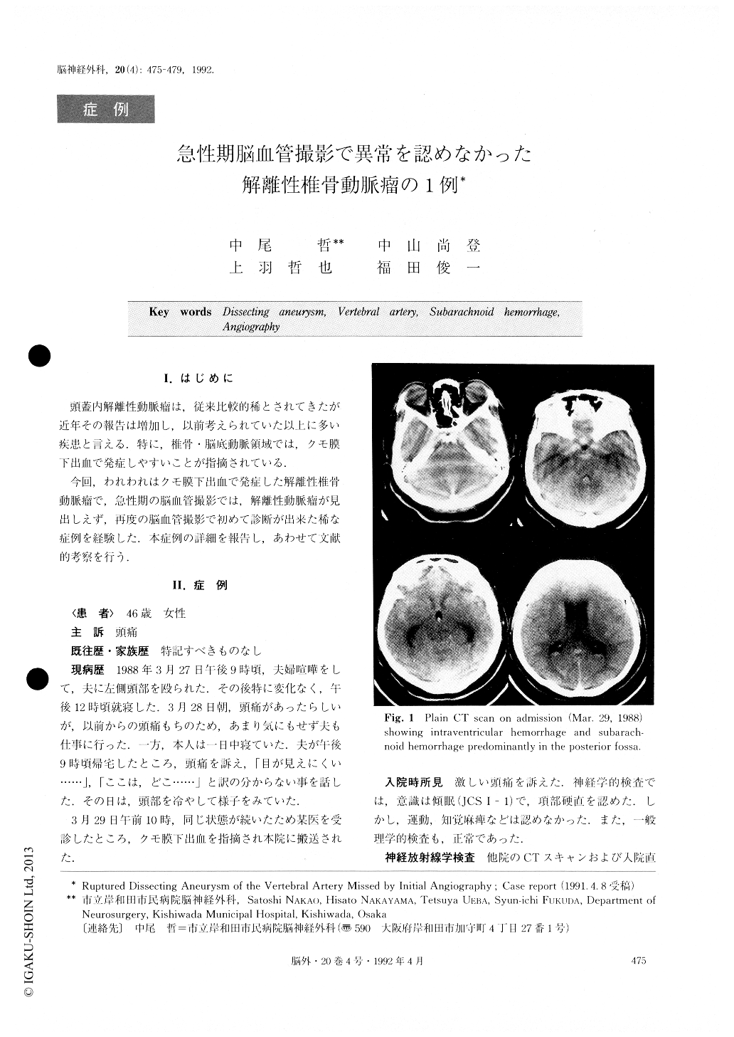
A 46-year-old woman had been well until she com-plained of headache. She was admitted to a local hos-pital and found to have SAH. She was transferred to our clinic for further examination. On admission the pa-tient was drowsy but able to he aroused. Her neurolo-gical state was normal except for a mildly stiff neck. Computed tomography (CT) demonstrated massive SAFI in the basal cistern and intraventricular hemor-rhage in all ventricles. CT also demonstrated acute hy-drocephalus. Four-vessel cerebral angiography was per-formed using transfemoral catheterization. No definite abnormalities, except for a small aneurysm in the caver-nous portion of the right internal carotid artery, were found. Because aneurysm in the cavernous portion could not cause SAH, we could not define the origin of the SAH. However, retrospectively, a slightly irregular wall of the left intracranial vertebral artery was evi-dent. Repeated angiography performed 28 days later re-vealed aneurysmal dilatation of this left vertebral artery. Double density of the contrast material was found in the aneurysmal dilatation. Retention of the contrast medium was also seen in the late capillary phase. From these angiographic findings, the aneurysm was diagnosed as being a dissecting aneurysm.
Direct surgical attack on the vertebral aneurysm was performed via a left suboccipital craniectomy. A fusi-form dilatation of the left vertebral artery showed that the dilated segment was blue, indicating subadventitial hematoma. It was clear that the aneurysm was not saccular but dissecting. Neither clipping of the aneurysm nor proximal clipping of the vertebral artery without occlusion of left posterior inferior cerebral artery was possible. Therefore, wrapping of the aneurysm was carried out.
The postoperative course was uneventful except for left hearing disturbance and transient hoarseness. There was no recurrence of SAI I in the following 3 years.
Copyright © 1992, Igaku-Shoin Ltd. All rights reserved.