Japanese
English
- 有料閲覧
- Abstract 文献概要
- 1ページ目 Look Inside
I.はじめに
頭蓋内解離性脳動脈瘤は約83%が椎骨脳底動脈系に発生し13),臨床像では内頸動脈系と比較してクモ膜下出血(SAH)で発症する症例が虚血病変例より多いとされている.また,再出血については嚢状動脈瘤より発症早期に起こりやすく,さらに再出血後の予後も不良であり外科的治療が必要と考えられている.しかし,直達手術(Trapping, Proximal Occlusionなど)3,7,12,14)や血管内手術川など治療法の選択も術後の合併症の問題もあり,未だ確立されていない3,6,12).
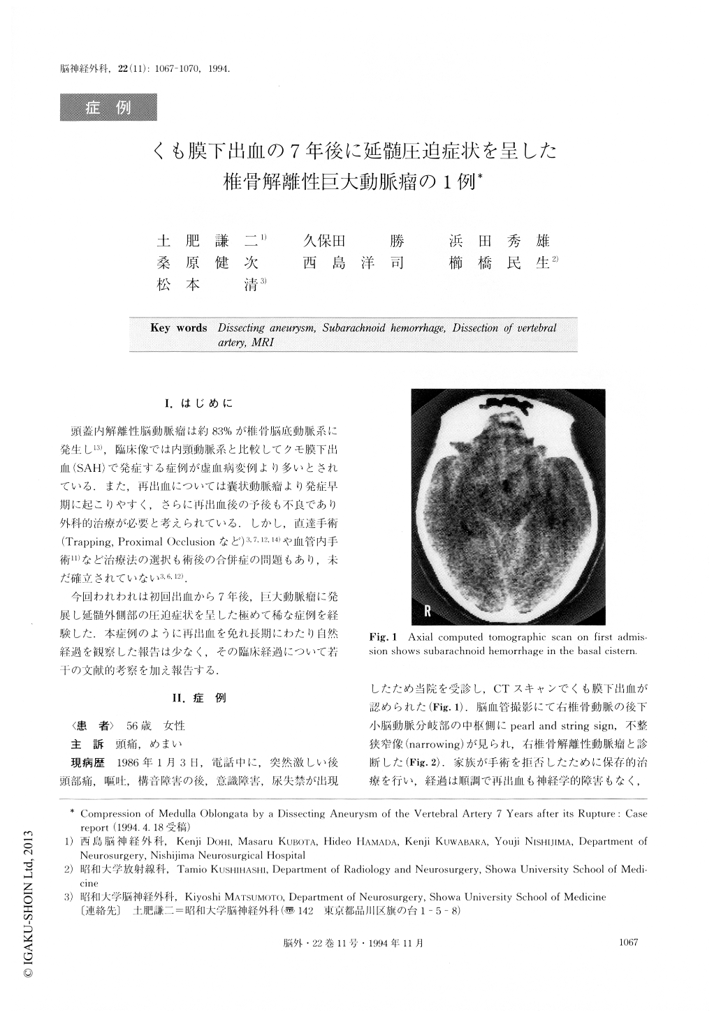
今回われわれは初回出血から7年後,巨大動脈瘤に発展し延髄外側部の圧迫症状を呈した極めて稀な症例を経験した.本症例のように再出血を免れ長期にわたり自然経過を観察した報告は少なく,その臨床経過について若干の文献的考察を加え報告する.
A 56-year-old female, who suffered a subarachnoid hemorrhage (SAH) due to spontaneous dissection of the right vertebral artery 7 years previously, was admit-ted to our hospital with headache and vertigo. She hadn't had any attacks of SAH for 7 years. A magnetic resonance imaging (MRI) showed a high signal intensi-ty mass and a low signal intensity due to calcification on the right ventrolateral surface of the medulla on both T1 and T2 weighted images. Vertebral angiogra-phy showed complete occlusion of the cervical segment of the right vertebral artery (VA). Left vertebral angio-graphy didn't reveal any retrograde filling of the intra-cranial segment of the right VA through VA union. Thus, the spontaneous entrapment by dissection of the vertebral artery was demonstrated 7 years after SAH with MRI and serial angiography.

Copyright © 1994, Igaku-Shoin Ltd. All rights reserved.


