Japanese
English
- 有料閲覧
- Abstract 文献概要
- 1ページ目 Look Inside
I.はじめに
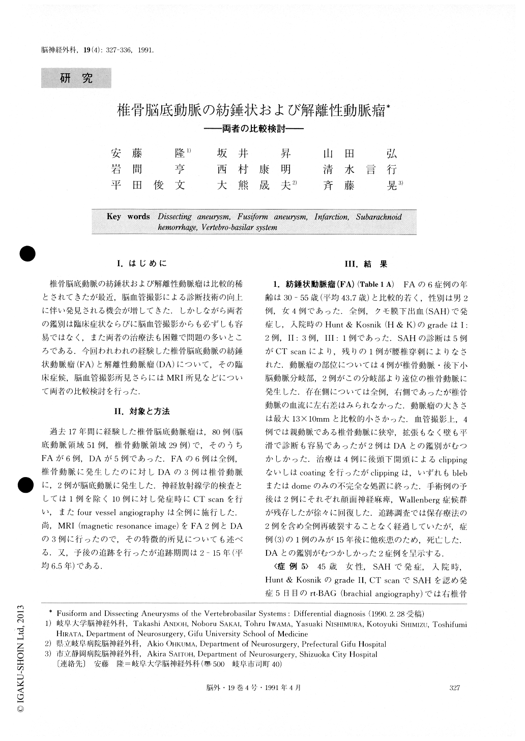
椎骨脳底動脈の紡錘状および解離性動脈瘤は比較的稀とされてきたが最近,脳血管撮影による診断技術の向上に伴い発見される機会が増してきた.しかしながら両者の鑑別は臨床症状ならびに脳血管撮影からも必ずしも容易ではなく,また両者の治療法も困難で問題の多いところである.今回われわれの経験した椎骨脳底動脈の紡錘状動脈瘤(FA)と解離性動脈瘤(DA)について,その臨床症候,脳血管撮影所見さらにはMRI所見などについて両者の比較検討を行った.
Abstract
Fusiform aneurysms (FA) and dissecting aneurysms (DA) of the vertebro-basilar systems have been thought to be rare, but recently reports of these aneurysms have increased. The differential diagnosis between FA and DA is requisite for deciding therapy or for prognosis, but it is often difficult to distinguish these aneurysms even if angiographies are conducted.The authors have treated 6 cases with FA and 5 cases with DA during the past 17 years.
As the initial symptom, subarachnoid hemorrhage (SAH) was noted in all 6 cases with FA. On the other hand, 2 cases of SAH, 2 cases of brain stem infarction, and 1 case of ischemic attack were noted in DA cases. The aneurysmal locations of FA were at the verte-bral artery (VA) in all 6 cases, and those of DA were at the VA in 3 cases and at the basilar artery (BA) in 2 cases.
As angiographic findings of DA, pearl & string signs were demonstrated in 3 cases, and retension of contrast medium was noted in 3 cases. Diagnosis of FA is com-paratively easy on angiography but when angiospasm exists, it is difficult to differentiate DA from FA. Con-sequently, repeated angiography is recommended. MR imaging findings of 2 cases with FA were compared with those of 3 cases with DA. No abnormal findings, excepting a dilatation of the signal-void area corres-ponding to the arterial blood flow were shown in FA, but various abnormalities were detected in all of the 3 cases with DA. Namely, an intimal flap and a double lumen in 1 case, intra-mural hematoma in 1 case, and a hematoma adjacent to the parent artery in 2 cases were demonstrated.
Thus, MR imaging was thought to be a useful means for distinguishing between FA and DA.

Copyright © 1991, Igaku-Shoin Ltd. All rights reserved.


