Japanese
English
- 有料閲覧
- Abstract 文献概要
- 1ページ目 Look Inside
I.はじめに
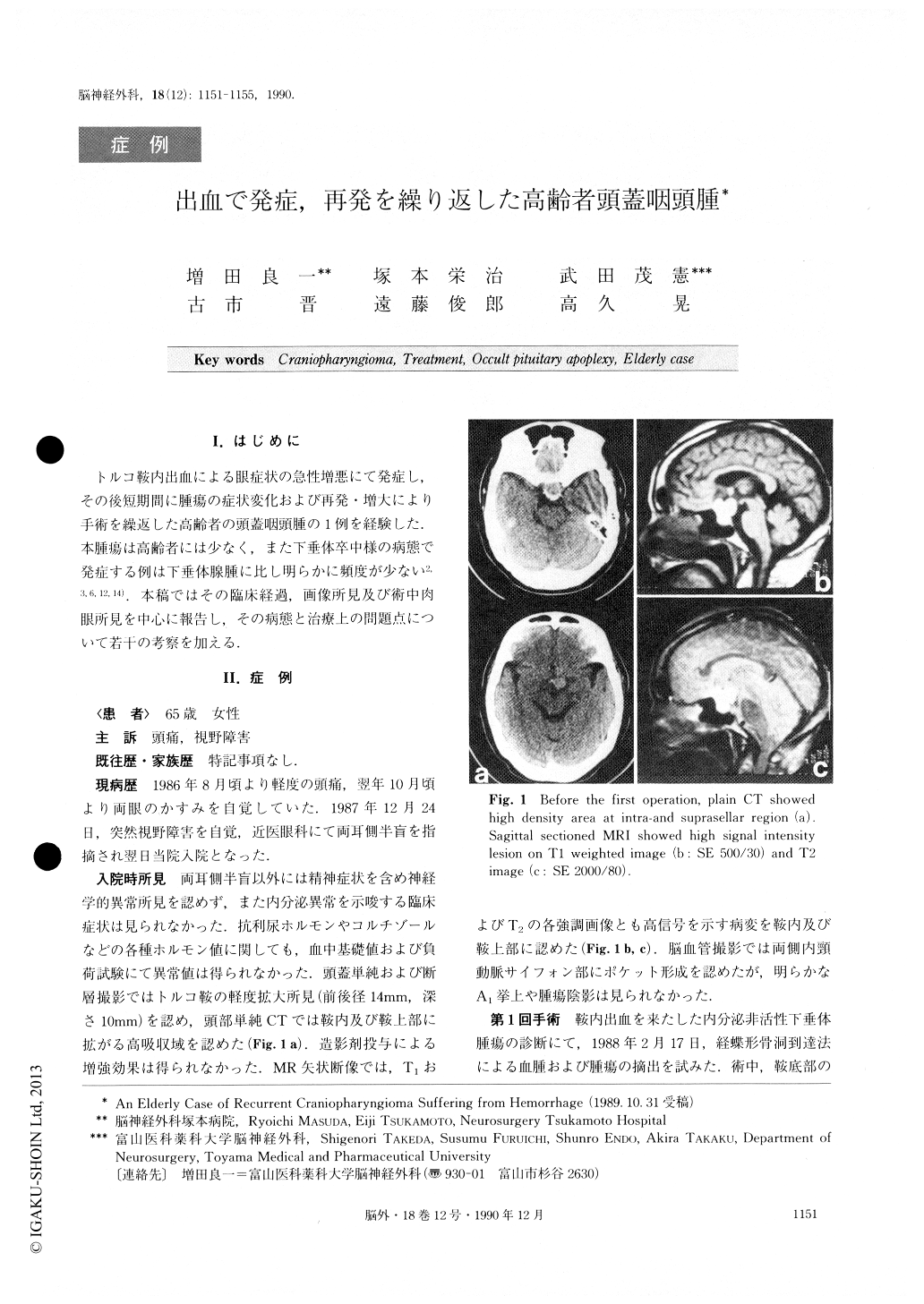
トルコ鞍内出血による眼症状の急性増悪にて発症し,その後短期間に腫瘍の症状変化および再発・増大により手術を繰返した高齢者の頭蓋咽頭腫の1例を経験した.本腫瘍は高齢者には少なく,また下垂体卒中様の病態で発症する例は下垂体腺腫に比し明らかに頻度が少ない2,3,6,12,14).本稿ではその臨床経過,画像所見及び術中肉眼所見を中心に報告し,その病態と治療上の問題点について若干の考察を加える.
A case of a 63-year-old female with craniopharyn-gioma is reported. She first suffered from occult pitui-tary apoplexy and had recurrent enlargement and char-acteristic changes of a tumor during short periods. This patient was hospitalized after suddenly developing hitemporal hemianopsia. An intra-and suprasellar hema-toma was revealed on computed tomography (CT) and magnetic resonance imaging (MRI). At the first opera-tion, the hematoma was removed totally by the trans-sphenoidal approach, but tumor tissues were not identi-fied. During the following 12 months, operations were repeated three times due to the recurrence and/or en-largement of a tumor associated with visual symptoms. Pathological diagnosis was squamous-type craniopharyn-gioma without any malignancy.
Microscopic appearance of the tumor apparently changed during the clinical course. The characteristic findings were revealed respectively on MRI and CI'.On the first preoperative MRI, a lesion of diffuse high signal intensity was observed on both Ti and T2 weighted images. At the second operation, the lesion was also revealed as having high signal intensity on Ti and T2 weighted images but the tumor had a large cyst with serous-yellowish fluid contents. A differential dia-gnosis was made on CT. At the third and fourth opera-tions, the tumor was solid and had atheromatous con-tents. The lesion was revealed as having low signal in-tensity on Ti image and high on T2 respectively.
Occult pituitary apoplexy with intra and suprasellar hemorrhage is very rare in cases of craniopharyngioma and only nine cases have been reported until now. It is also interesting with this high-aged patient that repe-ated recurrence and/or enlargement of a tumor with different microscopic appearances occurred during such short periods. The diagnostic significance of MRI and some points at issue for treatment in such cases were discussed.

Copyright © 1990, Igaku-Shoin Ltd. All rights reserved.


