Japanese
English
研究
CT像による頭蓋咽頭腫の治療評価
Treatment of Craniopharyngioma Estimated by Follow-up CT
久保田 紀彦
1
,
伊藤 治英
1
,
四十住 伸一
1
,
山本 信二郎
1
Toshihiko KUBOTA
1
,
Haruhide ITO
1
,
Shinichi AIZUMI
1
,
Shinjiro YAMAMOTO
1
1金沢大学脳神経外科
1Department of Neurosurgery, School of Medicine, University of Kanazawa
キーワード:
Craniopharyngioma
,
Follow-up CT
,
Cyst wall
,
Radiotherapy
Keyword:
Craniopharyngioma
,
Follow-up CT
,
Cyst wall
,
Radiotherapy
pp.1495-1501
発行日 1981年12月10日
Published Date 1981/12/10
DOI https://doi.org/10.11477/mf.1436201441
- 有料閲覧
- Abstract 文献概要
- 1ページ目 Look Inside
I.はじめに
CT scanの導入は,頭蓋内疾患の診断のみならず,その経過,ことに手術後の経過観察を容易にし,またCT scanによるfollow-upは術後の治療評価に必須のものとなった.
頭蓋咽頭腫は脳底部の脳神経,視床下部や脳幹,Willis環と密接しているため手術的に全摘することが困難な場合が少なくない4,11),このため手術療法に加えて種々な術後の補助療法が行われているが9),いまだ結論的なものはなく,厳重な追跡検査を必要とする.
Follow-up CT scans were taken from 12 cases of craniopharyngiomas after various treatment.
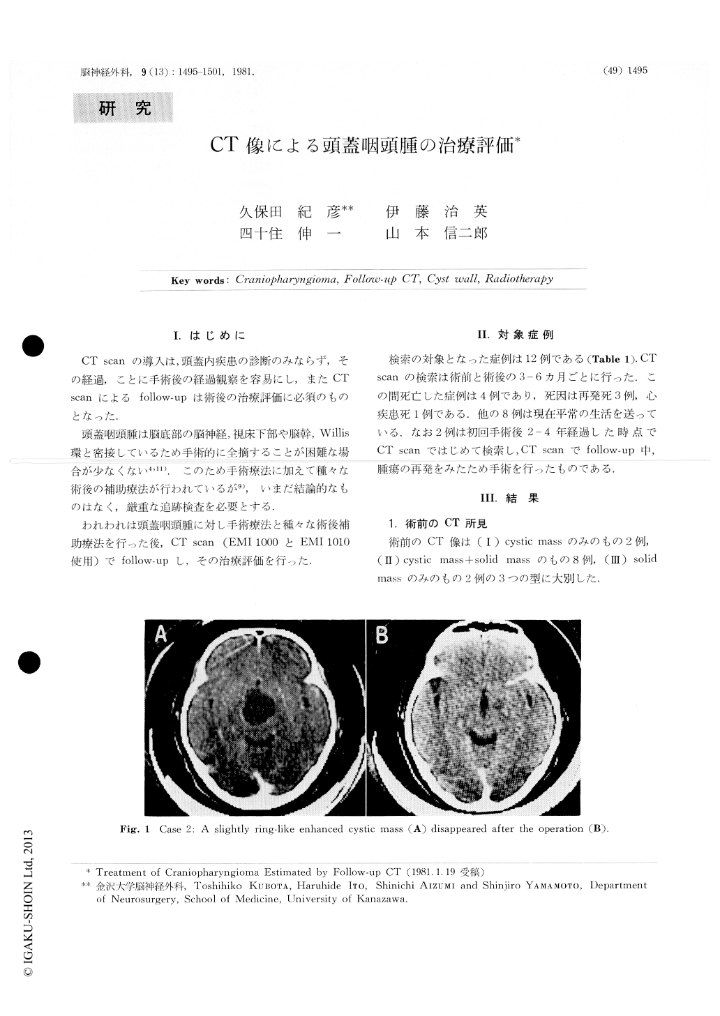
Preoperative CT findings of craniopharyngiomas could be classified into three types. Type 1 was a non-enhanced or a thinly ring-like enhanced large cystic mass. Type 2 was a thickly enhanced large cystic mass with small solid mass. Type 3 was a large solid mass.

Copyright © 1981, Igaku-Shoin Ltd. All rights reserved.


