Japanese
English
- 有料閲覧
- Abstract 文献概要
- 1ページ目 Look Inside
I.はじめに
血管撮影によって描出されない頭蓋内血管奇形をangiographically occult intracranial vascular malforma—tion(AOIVM)と呼び,頭痛,頭蓋内出血や難治性癲癇の原因として特にCT普及後多くの報告がなされている.AOIVMは,arteriovenous malformation, caver—nous angioma, capillary telangiectasia, venous angiomaへの分類5)が広く受け入れられている.
AOIVMの中には,診断に関して,脳内血腫,腫瘍(腫瘍内出血)等の鑑別が困難であるものがある.今回われわれは,術前検査で腫瘍内出血を否定しきれなかった,大脳基底核部の被膜形成された慢性脳内血腫の像を呈した静脈性血管腫の全摘出例を経験したので,診断,病理,手術方法について報告する.
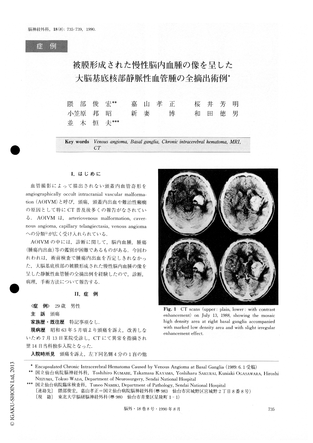
A case is reported of venous angioma at the right basal ganglia simulating the encapsulated chronic in-tracerebral hematoma. A 29-year-old man was admitted to our hospital on July 14, 1988 with a two-month his-tory of headache. Neurological examination revealed left homonymous lower quadrantic anopsia. CT scans showed a mosaic high density lesion at the right basal ganglia with extensive adjacent edema. MRI revealed that the high density lesion on CT scans was the corn-bination of a reticulated core of mixed signal intensity with a surrounding rim of decreased signal intensity. The lesion was accompanied with extensive edema. Followed up CT scans showed the transformation of the lesion and ring-shaped enhancement. A right fron-totemporal craniotomy was performed on August 9, 1988. After thorough dissection of the sylvian fissure and small corticotomy to the insula, a tough capsule was seen. There was blood in various stages of orga-nization in the capsule. A histological examination gave a diagnosis of venous angioma in the membrane similar to the outer membrane of chronic subdural hematomas. Postoperatively, the patient showed slight left motor weakness, but it gradually improved and he was dis-charged on foot, on October 19, 1988. There have been a lot of reports about angiographi-cally occult intracranial vascular malformation (AOIVM). But AOIVM at the basal ganglia is rare, and to our knowledge, only 8 cases have been reported. In our case, the presence of adjacent extensive edema, and ring-shaped enhancement on CT scans confused the preoperative diagnosis. Those findings might have been caused by encapsulation. By using CT scans and MRI, a complete and accurate diagnosis was impossi-ble. If a suitable operative approach is possible, we think more aggressive surgical procedures may be necessary to exclude underlying neoplasms and to avoid recurrent hemorrhages.

Copyright © 1990, Igaku-Shoin Ltd. All rights reserved.


