Japanese
English
- 有料閲覧
- Abstract 文献概要
- 1ページ目 Look Inside
I.はじめに
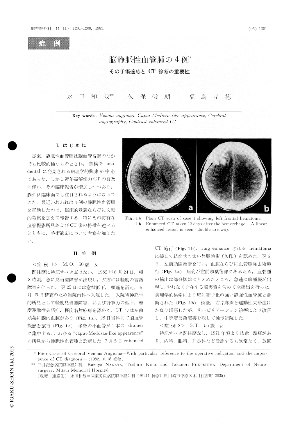
従来,静脈性血管腫は脳血管奇形のなかでも比較的稀なものとされ,剖検でinci-dentalに発見される病理学的興味が中心であった.しかし近年高解像力CTの普及に伴い,その臨床報告が増加しつつあり,脳外科臨床面でも注目されるようになってきた,最近われわれは4例の静脈性血管腫を経験したので,臨床的意義ならびに文献的考察を加えて報告する.特にその特有な血管撮影所見およびCT像の特徴を述べるとともに,手術適応について考察を加えたい.
Four cases of venous angioma, one cerebral andthree in the cerebellum, are reported.
Case 1. A 50-year-old woman who had a suddenattack of headache and disorientation was admitted tothe Mitsui Memorial Hospital. Neurological exami-nation revealed slight disorientation, mild motoraphasis and right hemiparesis. Plain CT scan onadmission showed a left frontal hematoma. Leftcerebral angiomas demonstrated a caput-Medusae-like lesion which consisted of numerous small veinsand drained into one single enlarged vein. EnhancedCT scan taken 12 days after the attack demonstrated alinear enhancement next of the hematoma.

Copyright © 1983, Igaku-Shoin Ltd. All rights reserved.


