Japanese
English
- 有料閲覧
- Abstract 文献概要
- 1ページ目 Look Inside
- 参考文献 Reference
Ⅰ.はじめに
もやもや病は,両側内頚動脈終末部に慢性進行性の狭窄を生じ,代償的に脳底部に異常血管網が形成される原因不明の疾患である11).一方,川崎病は乳児および幼児において原因不明の系統的血管炎を主体とする疾患であり6),活動期に稀ながら脳梗塞を合併する4,7,12,15,17).川崎病活動期のもやもや病合併の報告はないが,川崎病罹患歴のあるもやもや病の報告がある1,3,8,9,13,14).今回,川崎病の既往があるもやもや病3例を経験したので,文献的考察を交えて報告する.
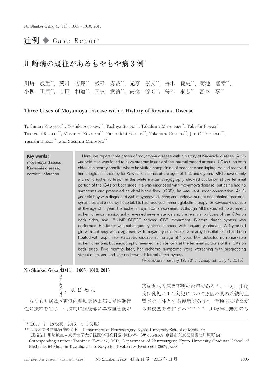
Here, we report three cases of moyamoya disease with a history of Kawasaki disease. A 33-year-old man was found to have stenotic lesions of the internal carotid arteries(ICAs)on both sides at a nearby hospital where he visited complaining of headache and lisping. He had received immunoglobulin therapy for Kawasaki disease at the ages of 1, 2, and 6 years. MRI showed only a chronic ischemic lesion in the white matter. Angiography showed occlusion at the terminal portion of the ICAs on both sides. He was diagnosed with moyamoya disease, but as he had no symptoms and preserved cerebral blood flow(CBF), he was kept under observation. An 8-year-old boy was diagnosed with moyamoya disease and underwent right encephaloduroarteriosynangiosis at a nearby hospital. He had received immunoglobulin therapy for Kawasaki disease at the age of 1 year. His ischemic symptoms worsened. Although MRI detected no apparent ischemic lesion, angiography revealed severe stenosis at the terminal portions of the ICAs on both sides, and 123I-IMP SPECT showed CBF impairment. Bilateral direct bypass was performed. His father was subsequently also diagnosed with moyamoya disease. A 4-year-old girl with epilepsy was diagnosed with moyamoya disease at a nearby hospital. She had been treated with aspirin for Kawasaki disease at the age of 1 year. MRI detected no remarkable ischemic lesions, but angiography revealed mild stenosis at the terminal portions of the ICAs on both sides. Five months later, her ischemic symptoms were worsening with progressing stenotic lesions, and she underwent bilateral direct bypass.

Copyright © 2015, Igaku-Shoin Ltd. All rights reserved.


