Japanese
English
- 有料閲覧
PPV購入案内
1,320円 (1,200円+税10%) こちらは、139ページから146ページの文献です。購入には医書.jp本会員登録が必要です。
会員登録完了後に改めて上記「購入サイトへ」を選択してください。
または「購入サイトへ」選択後に「購入ログイン」、「新規会員登録」の順に進んでください。
会員登録について詳しくはをご確認ください。
- Abstract 文献概要
- 1ページ目 Look Inside
- 参考文献 Reference
Ⅰ.はじめに
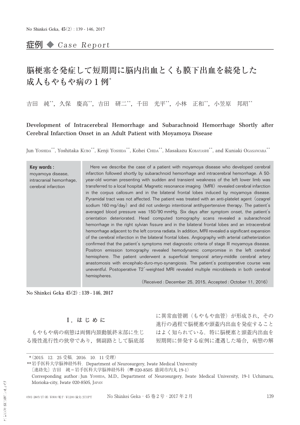
もやもや病の病態は両側内頚動脈終末部に生じる慢性進行性の狭窄であり,側副路として脳底部に異常血管網(もやもや血管)が形成され,その進行の過程で脳梗塞や頭蓋内出血を発症することはよく知られている.特に脳梗塞と頭蓋内出血を短期間に併発する症例に遭遇した場合,病態の解釈や治療の選択に苦慮する.成人もやもや病において,頭蓋内出血を発症してから短期間に脳梗塞を併発した症例は少なからず報告されている3,10,12,14,15).その病態としては,すでに脳血流が低灌流状態で脳血管予備能が低くなっているところに,出血による頭蓋内圧の変化,脱水,低血圧,血管攣縮,低炭酸ガス血症などが生じ,もやもや血管の側副血行路の流量低下を来すと考えられている15).一方,脳梗塞発症後に頭蓋内出血を来した成人もやもや病の症例は稀と考えられ,われわれが渉猟し得た範囲では1報告のみである1).今回われわれは,脳梗塞発症後に脳内出血とくも膜下出血を短期間に続発した成人もやもや病の1例を経験したので,若干の考察とともに報告する.
Here we describe the case of a patient with moyamoya disease who developed cerebral infarction followed shortly by subarachnoid hemorrhage and intracerebral hemorrhage. A 50-year-old woman presenting with sudden and transient weakness of the left lower limb was transferred to a local hospital. Magnetic resonance imaging(MRI)revealed cerebral infarction in the corpus callosum and in the bilateral frontal lobes induced by moyamoya disease. Pyramidal tract was not affected. The patient was treated with an anti-platelet agent(ozagrel sodium 160mg/day)and did not undergo intentional antihypertensive therapy. The patient's averaged blood pressure was 150/90mmHg. Six days after symptom onset, the patient's orientation deteriorated. Head computed tomography scans revealed a subarachnoid hemorrhage in the right sylvian fissure and in the bilateral frontal lobes and an intracerebral hemorrhage adjacent to the left corona radiata. In addition, MRI revealed a significant expansion of the cerebral infarction in the bilateral frontal lobes. Angiography with arterial catheterization confirmed that the patient's symptoms met diagnostic criteria of stage Ⅲ moyamoya disease. Positron emission tomography revealed hemodynamic compromise in the left cerebral hemisphere. The patient underwent a superficial temporal artery-middle cerebral artery anastomosis with encephalo-duro-myo-synangiosis. The patient's postoperative course was uneventful. Postoperative T2*-weighted MRI revealed multiple microbleeds in both cerebral hemispheres.
Copyright © 2017, Igaku-Shoin Ltd. All rights reserved.