Japanese
English
- 有料閲覧
- Abstract 文献概要
- 1ページ目 Look Inside
- 参考文献 Reference
Ⅰ.はじめに
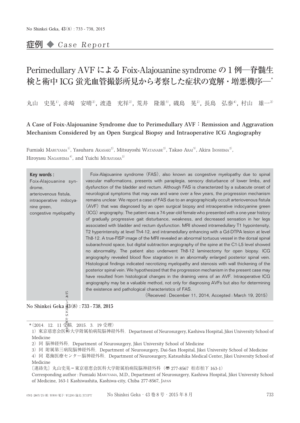
Foix-Alajouanine syndrome(FAS)は,亜急性に進行する対麻痺,下肢感覚障害,膀胱直腸障害を主症状とする症候群で,1926年にFoixらにより亜急性壊死性脊髄炎として報告された6).それ以降の症例検討でFASの病態は,脊髄硬膜動静脈瘻(dural arteriovenous fistula:dAVF),脊髄辺縁部動静脈瘻(perimedullary arteriovenous fistula:pAVF),脊髄内動静脈奇形(intramedullary arteriovenous malformation:iAVM)などの脊髄血管奇形(spinal vascular malformation:SVM)に伴うcongestive myelopathyであることが判明しており2,5,7-9),最終的には脊髄壊死を来すため症状は一般に不可逆的である4).しかし,FASにしばしば認められる「寛解・増悪を繰り返しながら段階的に進行していく症状」に関するメカニズムは十分な解析がなされておらず,未だ不明のままである.今回われわれは,MRI画像にてSVMが疑われたものの脊髄血管撮影検査では診断に至らず,脊髄生検と術中indocyanine green(ICG)蛍光血管撮影検査にて診断に至ったFASの1例を経験し,臨床症状,画像所見,手術所見などからFASの発症メカニズムについて検討したので報告する.
Foix-Alajouanine syndrome(FAS), also known as congestive myelopathy due to spinal vascular malformations, presents with paraplegia, sensory disturbance of lower limbs, and dysfunction of the bladder and rectum. Although FAS is characterized by a subacute onset of neurological symptoms that may wax and wane over a few years, the progression mechanism remains unclear. We report a case of FAS due to an angiographically occult arteriovenous fistula(AVF)that was diagnosed by an open surgical biopsy and intraoperative indocyanine green(ICG)angiography. The patient was a 74-year-old female who presented with a one-year history of gradually progressive gait disturbance, weakness, and decreased sensation in her legs associated with bladder and rectum dysfunction. MRI showed intramedullary T1 hypointensity, T2 hyperintensity at level Th4-12, and intramedullary enhancing with a Gd-DTPA lesion at level Th8-12. A true-FISP image of the MRI revealed an abnormal tortuous vessel in the dorsal spinal subarachnoid space, but digital subtraction angiography of the spine at the C1-L5 level showed no abnormality. The patient also underwent Th8-12 laminectomy for open biopsy. ICG angiography revealed blood flow stagnation in an abnormally enlarged posterior spinal vein. Histological findings indicated necrotizing myelopathy and stenosis with wall thickening of the posterior spinal vein. We hypothesized that the progression mechanism in the present case may have resulted from histological changes in the draining veins of an AVF. Intraoperative ICG angiography may be a valuable method, not only for diagnosing AVFs but also for determining the existence and pathological characteristics of FAS.

Copyright © 2015, Igaku-Shoin Ltd. All rights reserved.


