Japanese
English
- 有料閲覧
- Abstract 文献概要
- 1ページ目 Look Inside
- 参考文献 Reference
Ⅰ.は じ め に
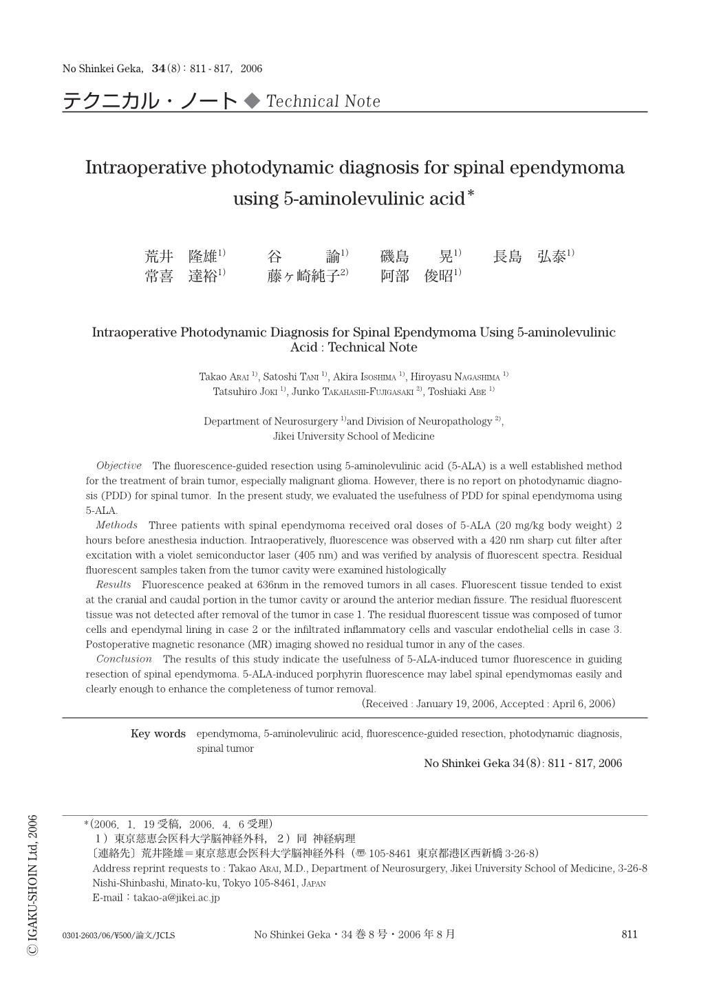
悪性脳腫瘍手術に対するphotodynamic diagnosis(PDD)は,近年急速に普及し重要な術中支援装置の1つとしてその地位を確立した.一方,脊髄腫瘍に対するPDDについてはいまだ報告されていない.今回われわれは,脊髄上衣腫におけるPDDの有用性を術中所見および病理組織学的所見をもとに検討した.
Objective The fluorescence-guided resection using 5-aminolevulinic acid (5-ALA) is a well established method for the treatment of brain tumor,especially malignant glioma. However,there is no report on photodynamic diagnosis (PDD) for spinal tumor. In the present study,we evaluated the usefulness of PDD for spinal ependymoma using 5-ALA.
Methods Three patients with spinal ependymoma received oral doses of 5-ALA (20 mg/kg body weight) 2 hours before anesthesia induction. Intraoperatively,fluorescence was observed with a 420 nm sharp cut filter after excitation with a violet semiconductor laser (405 nm) and was verified by analysis of fluorescent spectra. Residual fluorescent samples taken from the tumor cavity were examined histologically
Results Fluorescence peaked at 636nm in the removed tumors in all cases. Fluorescent tissue tended to exist at the cranial and caudal portion in the tumor cavity or around the anterior median fissure. The residual fluorescent tissue was not detected after removal of the tumor in case 1. The residual fluorescent tissue was composed of tumor cells and ependymal lining in case 2 or the infiltrated inflammatory cells and vascular endothelial cells in case 3. Postoperative magnetic resonance (MR) imaging showed no residual tumor in any of the cases.
Conclusion The results of this study indicate the usefulness of 5-ALA-induced tumor fluorescence in guiding resection of spinal ependymoma. 5-ALA-induced porphyrin fluorescence may label spinal ependymomas easily and clearly enough to enhance the completeness of tumor removal.

Copyright © 2006, Igaku-Shoin Ltd. All rights reserved.


