Japanese
English
- 有料閲覧
- Abstract 文献概要
- 1ページ目 Look Inside
I.はじめに
脊椎の血管腫は胸部X線写真(胸写)や胸腰椎単純写でincidentalに発見されることも少なくない.また剖検例の7-12%20)に認められ,無症状に経過していた血管腫がほとんどである.しかし脊髄症状を発現して直接的に手術が行われた症例報告は本邦では整形外科領域に散見される程度で,脳神経外科領域でも極めて少ない.この理由は椎体血管腫は極めて易出血性であり,術中止血のcontrolが困難なためである.もう一つは近年embolization3, 13)やradiation8, 27)による有効な治療結果が報告されているためでもある.
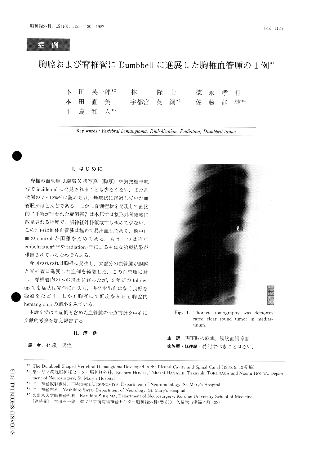
今回われわれは胸椎に発生し,大部分の血管腫が胸腔と脊椎管に進展した症例を経験した.この血管腫に対し,脊椎管内のみの摘出に終ったが,2年間のfollow—upでも症状は完全に消失し,再発や出血はなく良好な経過をたどり,しかも胸写にて軽度ながらも胸腔内hemangiomaの縮小をみている.
A 44-year-old male patient visited our clinic com-plaining of back pain since January, 1984 and feeling of weakness at the lower extremities, sensory and vesi-corectal disturbance since March of the same year. Hypesthesia and hypalgesia were observed at Th10 or below. The chest X-ray findings revealed an egg-sized tumor mass at the level of Th8. Metrizamide CT also demonstrated thoracic epidural mass. The charac-teristic findings of hemangioma showing delayed appearance of the tumor stain were also observed by angiography.

Copyright © 1987, Igaku-Shoin Ltd. All rights reserved.


