Japanese
English
症例
人工塞栓術中,腫瘍内およびくも膜下出血をきたしたのう胞性髄膜腫の1例
Cystic Meningioma associated with Intratumor and Subarachnoid Hemorrhage during Embolization: A casereport
正島 和人
1
,
林 隆士
1
,
宇都宮 英綱
2
,
森高 一彦
3
,
本田 英一郎
3
,
東原 英之
4
Kazuhito SHOJIMA
1
,
Takashi HAYASHI
1
,
Hidetsuna UTSUNOMIYA
2
,
Kazuhiko MORITAKA
3
,
Eiichiro HONDA
3
,
Hideyuki HIGASHIHARA
4
1聖マリア病院脳神経外科
2福岡大学放射線科
3久留米大学脳神経外科
4聖マリア病院神経放射線科
1Department of Neurosugery, St. Mary's Hospital
2Department of Radiology, Fukuoka University School of Medicine
3Department of Neurosurgery, Kurume University School of Medicine
4Department of Neuroradiology, St. Mary's Hospital
キーワード:
Cystic meningioma
,
Subarahnoid hemorrhage
,
Embolization
Keyword:
Cystic meningioma
,
Subarahnoid hemorrhage
,
Embolization
pp.919-924
発行日 1986年6月10日
Published Date 1986/6/10
DOI https://doi.org/10.11477/mf.1436202249
- 有料閲覧
- Abstract 文献概要
- 1ページ目 Look Inside
I.はじめに
のう胞性髄膜腫の発生頻度は髄膜腫の1.2-4.2%程度であるといわれ,稀とされている4,22).また,髄膜腫由来の腫瘍性頭蓋内出血は文献上稀である1,9,11,14,21).
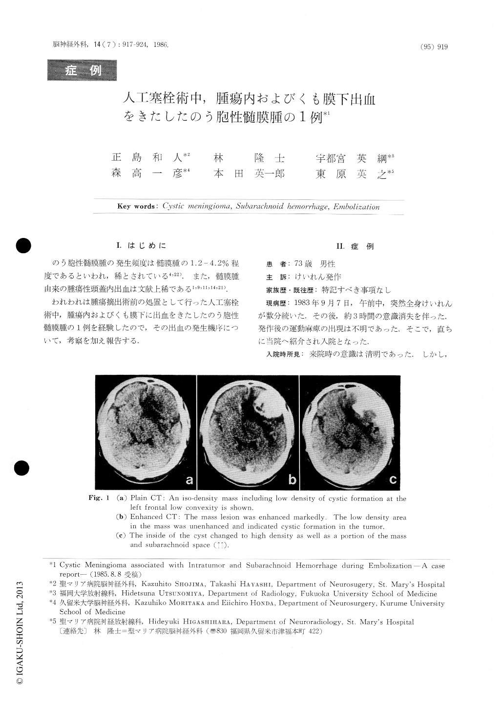
われわれは腫瘍摘出術前の処置として行った人工塞栓術中,腫瘍内およびくも膜下に出血をきたしたのう胞性髄膜腫の1例を経験したので,その出血の発生機序について,考察を加え報告する.
We present a case of a cystic meningioma accom-panied with hemorrhage in a cyst and adjacent subarachnoid space that occurred while preoperative embolization in feeders for the tumor was being applied.
A 73-year-old male patient was admitted for a complaint of convulsion. Under CT examination, a tumor was observed at the left frontal convexity and found to be fed by the middle cerebral artery shown in the left cerebral angiograms. The tumor was diagnosed meningioma.
After removing the tumor, we conducted histologi-cal study.

Copyright © 1986, Igaku-Shoin Ltd. All rights reserved.


