Japanese
English
- 有料閲覧
- Abstract 文献概要
- 1ページ目 Look Inside
I.はじめに
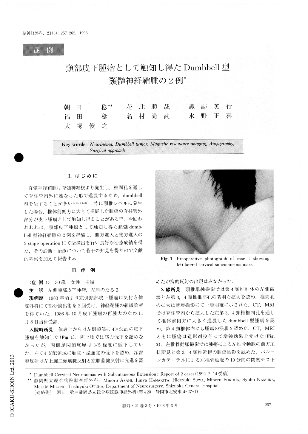
脊髄神経鞘腫は脊髄神経根より発生し,椎間孔を通して脊柱管内外に連なった形で進展するため,dumbbell型を呈することが多い1,13,14,21).特に頸椎レベルに発生した場合,椎体前側方に大きく進展した腫瘍の脊柱管外部分が皮下腫瘤として触知し得ることがある27).今回われわれは,頸部皮下腫瘤として触知し得た頸髄dumb—bell型神経鞘腫の2例を経験し,側方進入と後方進入の2stage operationにて全摘出を行い良好な治療成績を得た,その診断・治療について若干の知見を得たので文献的考察を加えて報告する.
Two cases of dummbell cervical neurinomas with massive subcutaneous extension were reported.
The first case was A 30-year-old woman who was admitted to our hospital because she had been aware of a left lateral cervical subcutaneous mass and was suf-fering from shoulder dullness. On admission, neurolo-gical examination revealed hypesthesia to touch and pain in the segmental area of C4, and hyperreflexia in the left biceps and patellar tendon reflexes. Plain X-ray showed enlargement of the left C3/4 intervertebral foramen. CT scan, post-myelogram CT and MRI de-monstrated a dumbbell shaped tumor at the level of C3 -4. Angiogram showed an anterior shift of the left ver-tebral artery (VA) and tumor stain. Temporary occlu-sion of the left VA by a balloon catheter was per-formed leaving no neurological deficits.
The second case was a 36-year-old woman who had been aware of a left lateral cervical subcutaneous mass. She complained of shoulder pain and finger clumsiness. On admission, neurological examination revealed weakness of the left deltoid muscle, hypesthesia in the segmental area of C3 - 4 and exaggeration of all deep tendon reflexes in the left-side extremities. Plain X-ray showed enlargement of the C2/3, C3/4 and C4/5 inter-verebral foramina. CT scan, post myelogram CT and MRI demonstrated a dumbbell shaped tumor at the level of C2 - 5. Angiogram showed an anteromedial shift of the left VA and tumor stain. Temporary occlu-sion test of the left VA by a balloon catheter was per-formed with negative results.
In each case two-stage operations were undertaken with excellent results. The lateral or anterolateral approach was used firstly to extirpate the extracanal portion of the tumor which extended anterolaterally through the intervertebral foramen. Secondly, removal of the intracanal and intradural portion of the tumor was performed, using the posterior approach. For the prevention of postoperative instability of the cervical spine, anterior fusion by iliac bone graft and immobi-lization by Haloshoulder brace were performed. In the second case the left VA was sacrificed during the op-erative procedures but no neurological deficits de-veloped postoperatively.
Copyright © 1993, Igaku-Shoin Ltd. All rights reserved.