Japanese
English
- 有料閲覧
- Abstract 文献概要
- 1ページ目 Look Inside
I.はじめに
下垂体inicroadenoinaの神経放射線学的診断は,従来thin-section tomographyによるトルコ鞍底lamina duraの変化によって診断されてきたが,腺腫直径が5mm以下のmicroadenomaでは診断が困難であった13,18).また最近high resolution CTの開発により,CTによる下垂体microadenomaの診断例が増加しているが,下垂体microadenomaのCT所見は造影前に,low dense,isodense,hyperdenseと種々のdensityを示し,造影後にhypodenseを示す場合5,15)と増強効果を示す場合5,9)があり,microadenomaに特有なCT所見はない.
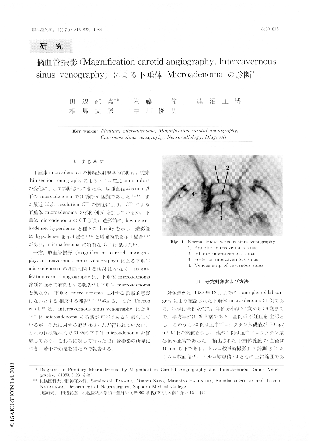
一方,脳血管撮影(magnification carotid angiography,intercavernous sinus venography)による下垂体microadenomaの診断に関する検討は少なく,magnification carotid angiographyは,下垂体microadenoma診断に極めて有効とする報告7)と下垂体macroadenomaと異なり,下垂体microadenomaに対する診断的意義はないとする相反する報告2,11,12)がある.
Carotid angiography was performed on 12 cases of pituitary microadenomas at 2.3×magnified frontal projection and 3.0×magnified lateral projection. Magnification carotid angiograms were analyzed to determine the pathologic findings in 12 cases on 23 sides. Carotid angiograms demonstrated compressed posterior pituitary gland in 3 of 12 cases and 4 of 23 sides which resulted from the expansion of pituitary microadenomas. But carotid angiogram failed to demonstrate any evidence of tumor stain, abnormal capsular arteries or hypertrophied inferior hypophyseal arteries.

Copyright © 1984, Igaku-Shoin Ltd. All rights reserved.


