Japanese
English
- 有料閲覧
- Abstract 文献概要
- 1ページ目 Look Inside
I.はじめに
下垂体microadenomaに対し筆者らは過去数年間にわたりthin-section hypocycloidal tomography5),inter—cavernous sinus venography8),high resolution CT9)等を用いた診断の可能性と限界を検討し,それぞれにおける診断基準を明かにしてきた.その結果,high resolu—tion CTが最も有用であると思われたが,これを用いた場合でもmicroadenomaに対する診断率は約70-80%程度に留まり,とくに直径7mm以下の腺腫では重要な診断の手掛かりとなる下垂体茎の偏位・鞍隔膜の挙上等の所見はみられず,さらに直径5mm以下の腺腫では下垂体腺腫の存在を示すhypodensityを描出することさえも困難であった.
そこで,直径5mm以下のmicroadenomaのより精度の高い診断法の確立を目指してDynamic CTを行い,その所見を腺腫の存在を直接示すdirect signと腺腫による下垂体組織の偏位を示すindirect signに分けて検討した.
The advantage of high resolution CT in the diagno-sis of pituitary microadenomas has been established,but the diagnosis becomes more difficult when the pituitary microadenoma is less than 5mm in diameter. We have studied the usefulness of dynamic CT scans particularly for diagnosis of small microadenomas.
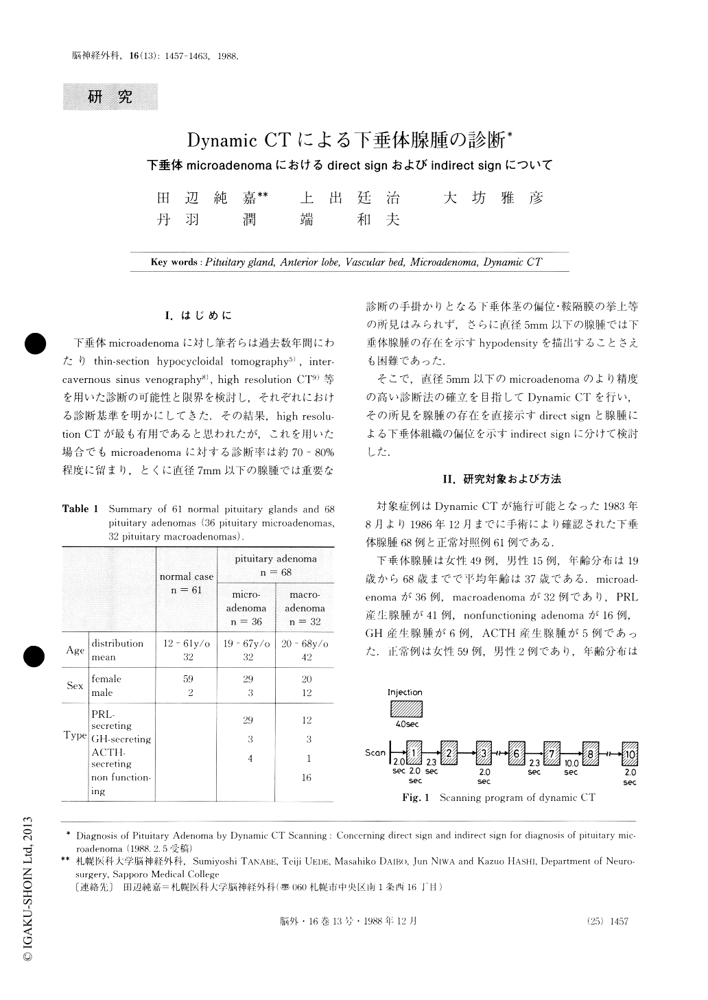
The dynamic CT scans were performed for 61 nor-mal pituitary glands and 68 pituitary adenomas (36 mic-roadenomas, 32 macroadenomas) with a GECT/T 9800 scanner. Coronal sections of 1.5mm thickness were taken at the plane just in front of the pituitary stalk of the pituitary gland.

Copyright © 1988, Igaku-Shoin Ltd. All rights reserved.


