Japanese
English
- 有料閲覧
- Abstract 文献概要
- 1ページ目 Look Inside
I.はじめに
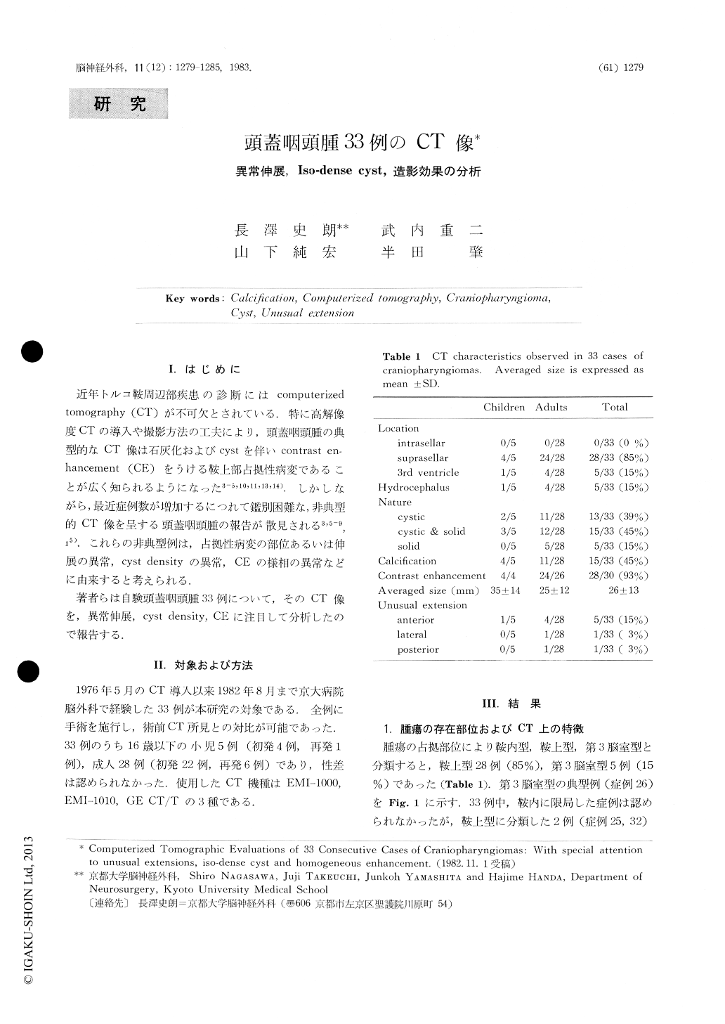
近年トルコ鞍周辺部疾患の診断にはcomputerizedtomography(CT)が不可欠とされている.特に高解像度CTの導入や撮影方法の工夫により,頭蓋咽頭腫の典型的なCT像は石灰化およびcystを伴いcontras en-hancement(CE)をうける鞍上部占拠性病変であることが広く知られるようになった3-5,10,11,13,14).しかしながら,最近症例数が増加するにつれて鑑別困難な,非典型的CT像を呈する頭蓋咽頭腫の報告が散見される3,5-9,15).これらの非典型例は,占拠性病変の部位あるいは伸展の異常,cyst densityの異常,CEの様相の異常などに由来すると考えられる.
著者らは自験頭蓋咽頭腫33例について,そのCT像を,異常伸展,cyst density,CEに注目して分析したので報告する.
Although craniopharyngiomas are widely known toexhibit three basic CT characteristics;calcification,cyst (s) and contrast enhancement (CE), several caseswith atypical CT manifestations have been reportedlately. These atypical manifestations can beclassifiedinto unusual extensions of the tumors, high densecyst and marked homogeneous CE. The CT scansobtained in our recent series of 33craniopharyngiomaswere evaluated to analyze tumoral extensions, cystdensity and CE.

Copyright © 1983, Igaku-Shoin Ltd. All rights reserved.


