Japanese
English
- 有料閲覧
- Abstract 文献概要
- 1ページ目 Look Inside
I.はじめに
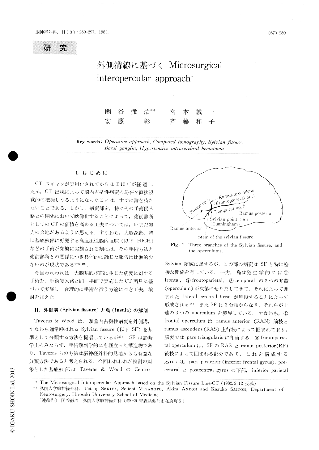
CTスキャンが実用化されてからほぼ10年が経過したが,CT出現によって脳内占拠性病変の局在を直接視覚的に把握しうるようになったことは,すでに論を待たないことである.しかし,病変部を,特にその手術侵入路との関係において映像化することによって,術前診断としてのCTの価値を高める工夫については,いまだ努力の余地があるように思える.すなわち,大脳深部,特に基底核部に好発する高圧性脳内血腫(以下HICH)などの手術が頻繁に実施される割には,その手術方法と術前診断との関係につき具体的に論じた報告は比較的少ないのが現状である4-6,19).
今回われわれは,大脳基底核部に生じた病変に対する手術を,手術侵入路と同一平面で実施したCT所見に基づいて実施し,合理的に手術を行う方途につき工夫,検討を加えた.
Since the advent of computed tomography (CT), it has become possible to localize and approach the intracranial mass, much more precisely and accurately than before.
Although it is needless to say that neurosurgeons should make efforts to minimize the brain damage accompanying surgical procedures, this principle especially applicable in the surgery for lesions deep in the cerebrum, such as the basal ganglia.
We discussed about operative techniques for the lesions of the basal ganglia, such as hypertensive intracerebral hematornas and operable brain tumor in this region, based on our special CT technique.

Copyright © 1983, Igaku-Shoin Ltd. All rights reserved.


