Japanese
English
- 有料閲覧
- Abstract 文献概要
- 1ページ目 Look Inside
I.はじめに
頭蓋内における間葉性混合腫瘍は,血管やくも膜および硬膜の線維芽細胞を主な発生母細胞とし,かつ粘液,骨あるいは脂肪成分等を有する16)ため多彩な組織像を呈することで知られている.また線維芽細胞腫は,線維性髄膜腫と発生部位において極めて近似しているにもかかわらず,その報告は意外に少ない1-5,7,8,10,11,17).
著者らは,左側頭頭頂部の硬膜の線維芽細胞を母地として発生したと思われる巨大な線維粘液腫の1治験例を経験したので,臨床病理学的検討を加えて報告する.
A 19-year-old, right handed female was admitted to Department of Neurosurgery in our hospital with paroxymal headache, nausea, vomiting and loss of concentration in January, 1975.
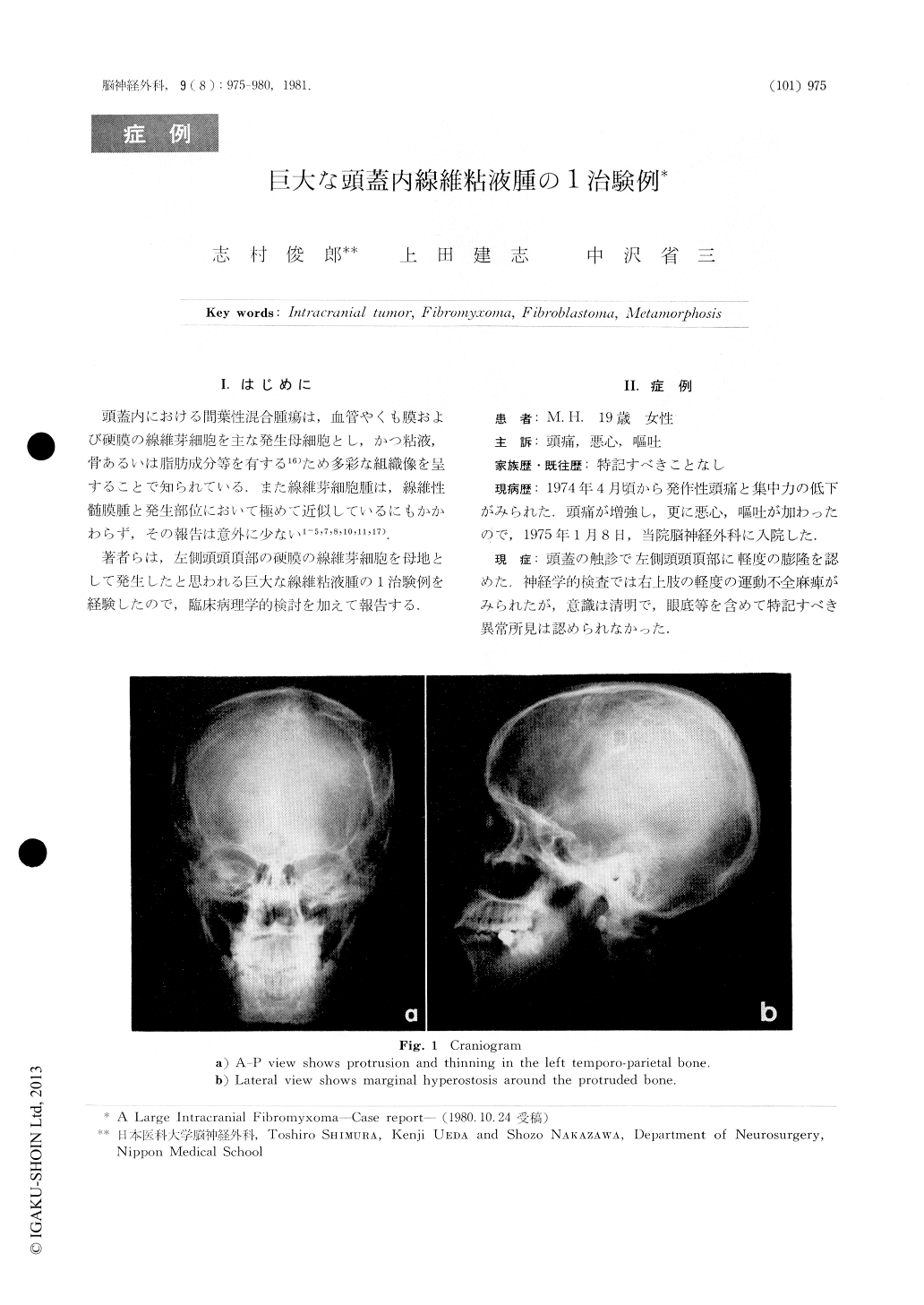
Neurological examination revealed slight motor weakness of the right upper extremities. The craniogram showed protrusion of thinned bone with marginal hyperostosis (measured 8.0×7.0cm in dimention) in the left temporoparietal region (Figs. 1a, b). The cerebral angiogram revealed a less vascular mass in the suprasylvian region (Figs. 2 a, b). The EEG findings showed polymorphous slow waves in the left temporo-parietal region.

Copyright © 1981, Igaku-Shoin Ltd. All rights reserved.


