Japanese
English
- 有料閲覧
PPV購入案内
1,320円 (1,200円+税10%) こちらは、267ページから271ページの文献です。購入には医書.jp本会員登録が必要です。
会員登録完了後に改めて上記「購入サイトへ」を選択してください。
または「購入サイトへ」選択後に「購入ログイン」、「新規会員登録」の順に進んでください。
会員登録について詳しくはをご確認ください。
- Abstract 文献概要
- 1ページ目 Look Inside
I.はじめに
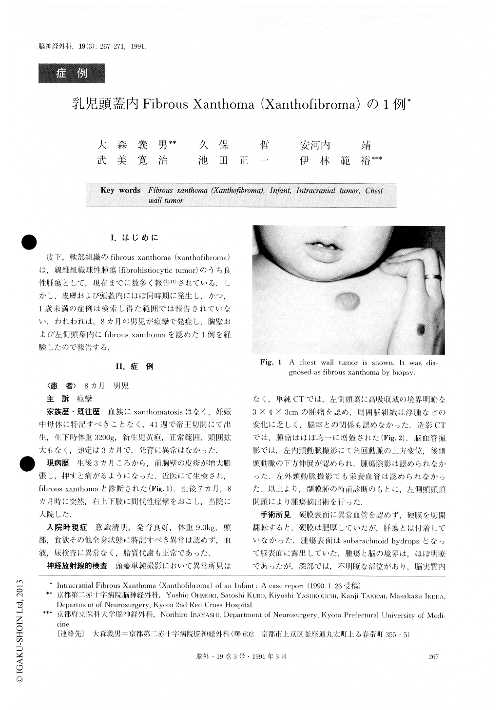
皮下,軟部組織のfibrous xanthoma(xanthofibroma)は,線維紐織球性腫瘍(fibrohistiocytic tumor)のうち良性腫瘍として,現在までに数多く報告11)されている.しかし,皮膚および頭蓋内にほぼ同時期に発生し,かつ,1歳未満の症例は検索し得た範囲では報告されていない.われわれは,8カ月の男児が痙攣で発症し,胸壁および左側頭葉内にfibrous xanthomaを認めた1例を経験したので報告する.
Abstract
A case of intracranial fibrous xanthoma (xanthofibro-ma) is reported. Intracranial fibrous xanthoma in infan-cy under the age of 1 year is extremely rare. This pa-tient was a 8-month-old boy with a history of convul-sive seizure. He had a previously known chest wall tumor which was diagnosed as fibrous xanthoma of the skin. Plain CT scan revealed a well defined high den-sity area in the left temporal lobe. The area was well enhanced with contrast media. At operation, it was found that the tumor did not attach to Jura mater and was almost well demarcated. Total removal of the tumor was performed. The patient has been doing well for these 6 months following craniotomy, with no sings of recurrence and no neurological deficits. Histologi-cally, the tumor was composed of fibrolastic cells and foamy phagocytic cells in storiform pattern. Some mul-tinucleated giant cells were found. Immunohistochemi-stry technique revealed that the tumor cells were nega-tive for GFAP, positive for Vimentin, positive for S-100 protein and negative for EMA. Our studies support the diagnosis of intracranial fibrous xanthoma coexistent with the same tumor found in the subcutaneous space of the chest wall of a boy under 1 year of age. We re-gard it as a rare incidence. Differential diagnosis and the characteristics of fibrous xanthoma were discussed.
Copyright © 1991, Igaku-Shoin Ltd. All rights reserved.