Japanese
English
- 有料閲覧
- Abstract 文献概要
- 1ページ目 Look Inside
I.はじめに
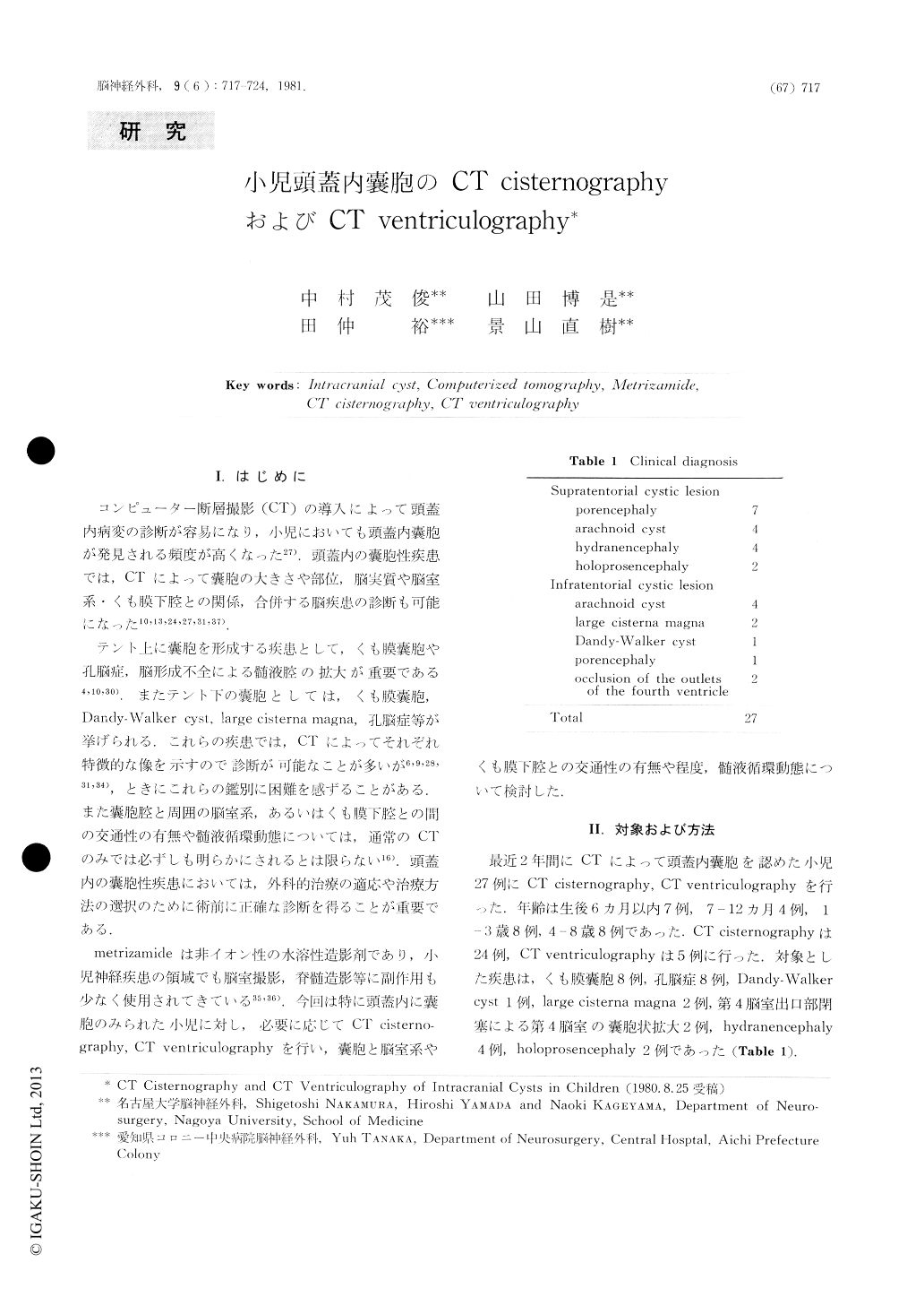
コンピューター断層撮影(CT)の導入によって頭蓋内病変の診断が容易になり,小児においても頭蓋内嚢胞が発見される頻度が高くなった27).頭蓋内の嚢胞性疾患では,CTによって嚢胞の大きさや部位,脳実質や脳室系・くも膜下腔との関係,合併する脳疾患の診断も可能になった10,13,24,27,31,37).
テント上に襲胞を形成する疾患として,くも膜嚢胞や孔脳症,脳形成不全による髄液腔の拡大が重要である4,10,30).またテント下の嚢胞としては,くも膜嚢胞,Dany-Walker cyst, large cisterna magna,孔脳症等が挙げられる.これらの疾患では,CTによってそれぞれ特徴的な像を示すので診断が可能なことが多いが6,9,28,31,34),ときにこれらの鑑別に困難を感ずることがある.また嚢胞腔と周囲の脳室系,あるいはくも膜下腔との間の交通性の有無や髄液循環動態については,通常のCTのみでは必ずしも明らかにされるとは限らない16).頭蓋内の嚢胞性疾患においては,外科的治療の適応や治療方法の選択のために術前に正確な診断を得ることが重要である.
Development of computerized tomography (CT) has greatly improved the ease and accuracy of identification of fluidcontaining intracranial cavities. CT reveals the size, location of the cyst, associated brain disorders and relationship between the cyst and the subarachnoid space or the ventricular system. An accurate diagnosis by plain CT is sometimes difficult to obtain. It is important to determine whether the cysts communicate with the ventricular system or subarachnoid space for the selection of the surgical treatment.

Copyright © 1981, Igaku-Shoin Ltd. All rights reserved.


