Japanese
English
- 有料閲覧
- Abstract 文献概要
- 1ページ目 Look Inside
1.序言
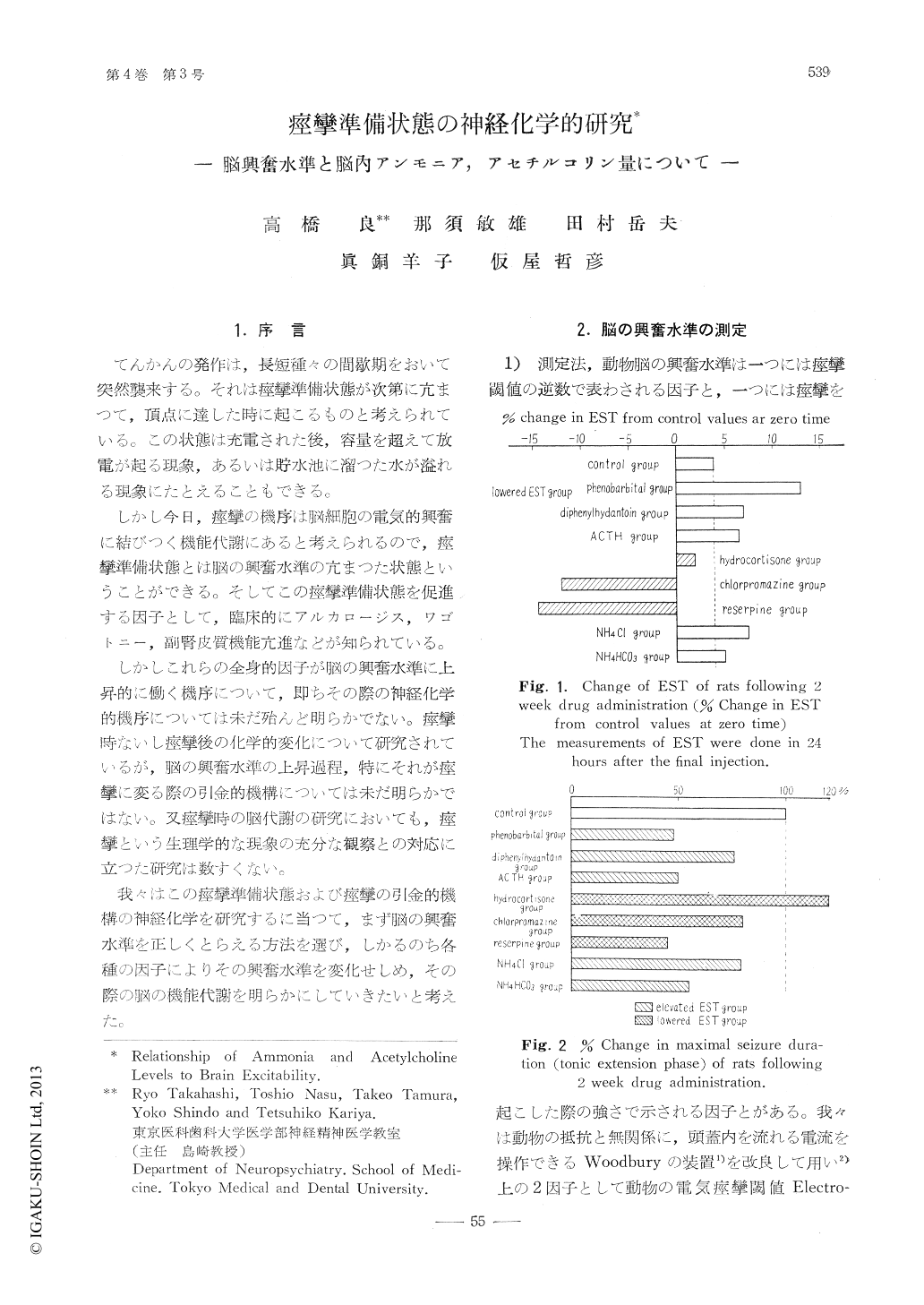
てんかんの発作は,長短腫々の間歇期をおいて突然襲来する。それは痙攣準備状態が次第に亢まつて,頂点に達した時に起こるものと考えられている。この状態は充電された後,容量を超えて放電が起る現象,あるいは貯水池に溜つた水が溢れる現象にたとえることもできる。
しかし今日,痙攣の機序は脳細胞の電気的興奮に結びつく機能代謝にあると考えられるので,痙攣準備状態とは脳の興奮水準の亢まつた状態ということができる。そしてこの痙攣準備状態を促進する因子として,臨床的にアルカロージス,ワゴトニー,副腎皮質機能亢進などが知られている。
The neurochemical mechanism concerned inchanging brain excitability are of importance forthe understanding of seizure pathophysiology.
I. Changes in rat brain excitability followingtwo week drug administration were measuredusing the electroshock seizure threshold (EST)and maximal seizure duration (MES) techniques,by modified Woodbury's apparatus.
It was noted that the EST was elevated in theanimals given phenobarbital, diphenylhydantoinand ACTH and lowered in the chlorpromazine.reserpine and hydrocortisone groups.
The duration of the tonic extension phase wasprolonged in the hydrocortisone group and short-ened in the other five groups.
Free ammonia and acetylcholine levels followingtwo week drug administration were measuredrespectively by Richter and Dawson's method andCrossland's method using the liquid air fixationtechnique.
Brain ammonia was increased in the hydrocorti-sone group, and decreased in the reserpine group.No significant change occured in the other fourgroups. Total acetylcholine was increased in thehydrocortisone group, and decreased in the reser-pine group, with no significant change occurringin the other four groups.
Brain ammonia levels did not correlate withEST changes. However there was a correlationbetween increased acetylcholine and increased du-ration of tonic extension phase (MES) in thehydrocortisone group.
In the reserpine group both the acetylcholinelevel and the MES were decreased, Acetylcholineis felt to influence the intensity of the seizurerather than the seizure threshold.
II. Brain ammonia and acetylcholine levels ofanimals in the course of the refractory period fol-lowing the maximal electroshock seizure (NIES)were measured using the liquid air fixation tech-nique.
As a measure of the refractory period of animalswas used the recovery time (RT50), which is, ac-cording to Woodbury, defined as the time requir-ed, after an initial electroshock seizure, for 50%of animals to recover to the extent that a secondelectroshock seizure can be elicited. The RT50for MES of adult d-d strain mice was 84 seconds(95% confidence limits were 77sec.-94sec.) andRT50 for EST seizure was 15.1 minutes (95%confidence limits 10min.-22.6min.).
Brain acetylcholine levels of mice during thefuror and the minimal seizure decreased by 30%compared with the acetylcholine level at rest. Itdecreased rapidly at the tonic flexion phase (TF)of MES, which lasted for 1 second after the elec-tric stimulation. But that decrease did not pro-gress in the tonic extension (TE) and clonic phase(Cl) of MES which lasted for 16 sec. followingTF, and increased toward the normal level. Itwas noteworthy that the acetylcholine reached thenormal level at about 80 seconds, which is theRT50 for MES, after the stimulation.
The brain ammonia increased steady followingthe stimulation, reached maximum level at the endof clonic phase and did not return to the normallevel in the RT50 for MES, but, in 15 minutes,RT50 for EST.
These facts support the above conclusion thatnot ammonia, but acetylcholine level correlateswith the duration of MES rather than the EST.Brain acetylcholine level of mice in the time offuror and minimal seizure is almost equal to thelevel at the TE or Cl phase of MES.
Animals given phenobarbital or diphenylhy-flantoin did not show TF and TE phase of MESand did not show any decrease of acetylcholineat 1sec. after the stimulation. At the Cl phaseof such treated mice, acetylcholine decreased tothe same degree as TE phase of non treatedanimals.
Therefore acetylcholine volume which is releas-ed in one sec after the stimulation, namely theacetylcholine releasing rate seems to determinethe seizure pattern.
Mice treated with EEP could give rise to com-plete maximal seizure following the stimulation,but did not show brain Ach decrease during theTF and TE phase. In this case, free acetylcholinehas seemed to accumulate in the brain.
Thus, not the rate, at which free Ach is de-stroyed by cholineesterase, but the rate at whichinactive bound acetylcholine is released to activefree form, is felt to be important for modulatingthe seizure intensity.
Copyright © 1960, Igaku-Shoin Ltd. All rights reserved.