Japanese
English
- 有料閲覧
- Abstract 文献概要
- 1ページ目 Look Inside
はじめに
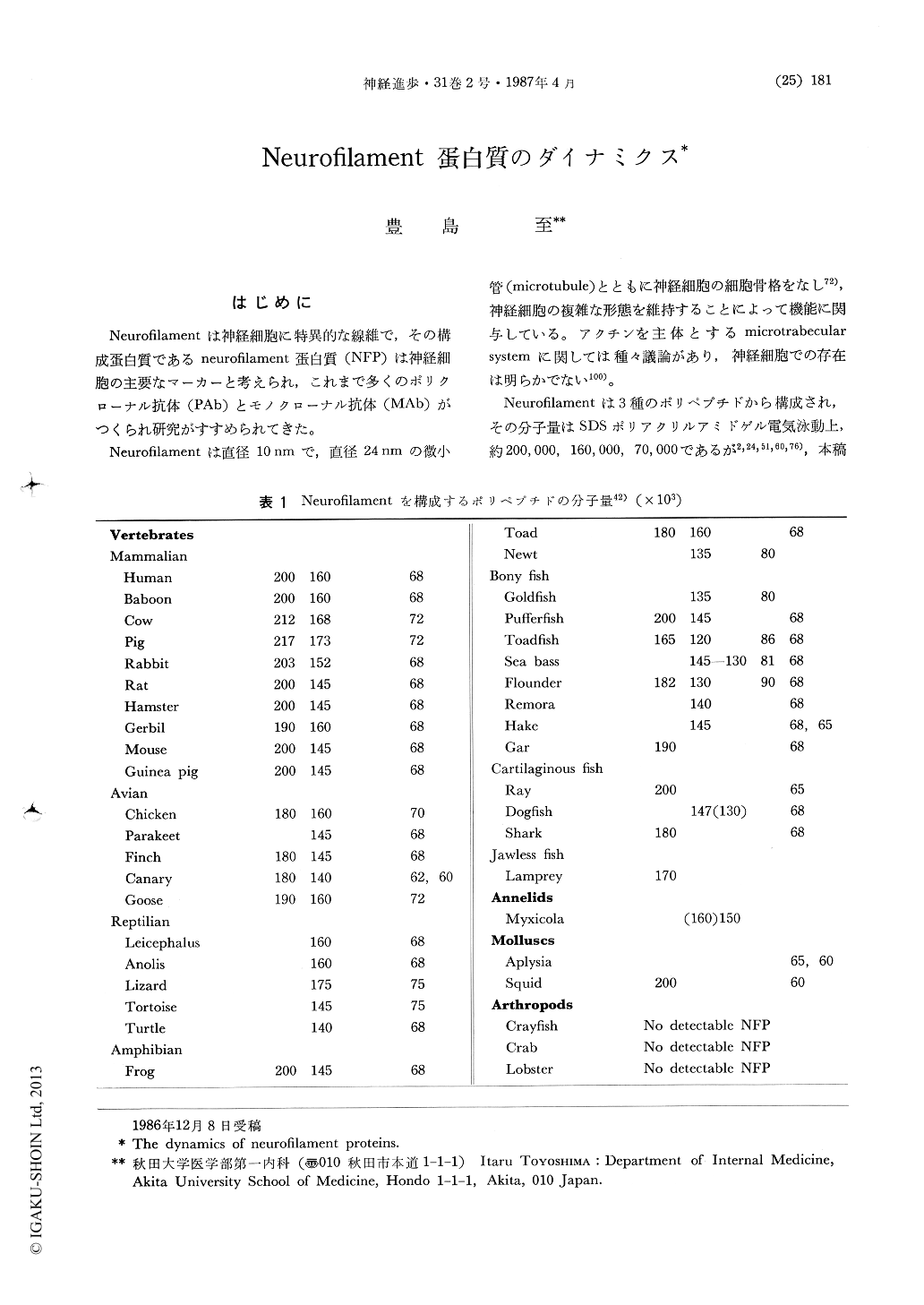
Neurofilamentは神経細胞に特異的な線維で,その構成蛋白質であるneurofilament蛋白質(NFP)は神経細胞の主要なマーカーと考えられ,これまで多くのポリクローナル抗体(PAb)とモノクローナル抗体(MAb)がつくられ研究がすすめられてきた。
Neurofilamentは直径10nmで,直径24nmの微小管(microtubule)とともに神経細胞の細胞骨格をなし72),神経細胞の複雑な形態を維持することによって機能に関与している。アクチンを主体とするmicrotrabecular systemに関しては種々議論があり,神経細胞での存在は明らかでない100)。
This review focused on the polyclonal and monoclonal antibodies (PAbs and MAbs) distinguishing the regional differences of neurofilament proteins (NFPs) in neurons.
Axonal neurofilaments consist of three peptides with high, middle and low molecular mass (NFP-H, M and L). All PAbs to each peptide stained most of axons but some of PAb to NFP-H or M failed to stain perikarya and dendrites of large neurons such as Purkinje cells in cerebellum and pyramidal cells in cerebral cortex. Our experimental results suggest that the differences are due to the different cross reactivities of these antibodies to perikaryal NFP-H and M. Perikarya of bovine spinal ganglion were dissected from freeze-dried sections and analysed by two-dimensional gel electrophoresis and immunoblotting with PAbs. Perikaryal NFP-H and M are more alkaline and apparent lower molecular mass with high heterogeneity compared to axonal NFP-H and M (Fig. 1). These differences have been revealed to be due to the different degree of phosphorylation which can be recognized by some series of MAbs to NFPs. Some MAb to non-phosphorylated NFP stained perikarya and dendrites but some other MAbs to phosphorylated NFP reacted to axonal type neurofilaments.

Copyright © 1987, Igaku-Shoin Ltd. All rights reserved.


