Japanese
English
- 有料閲覧
- Abstract 文献概要
- 1ページ目 Look Inside
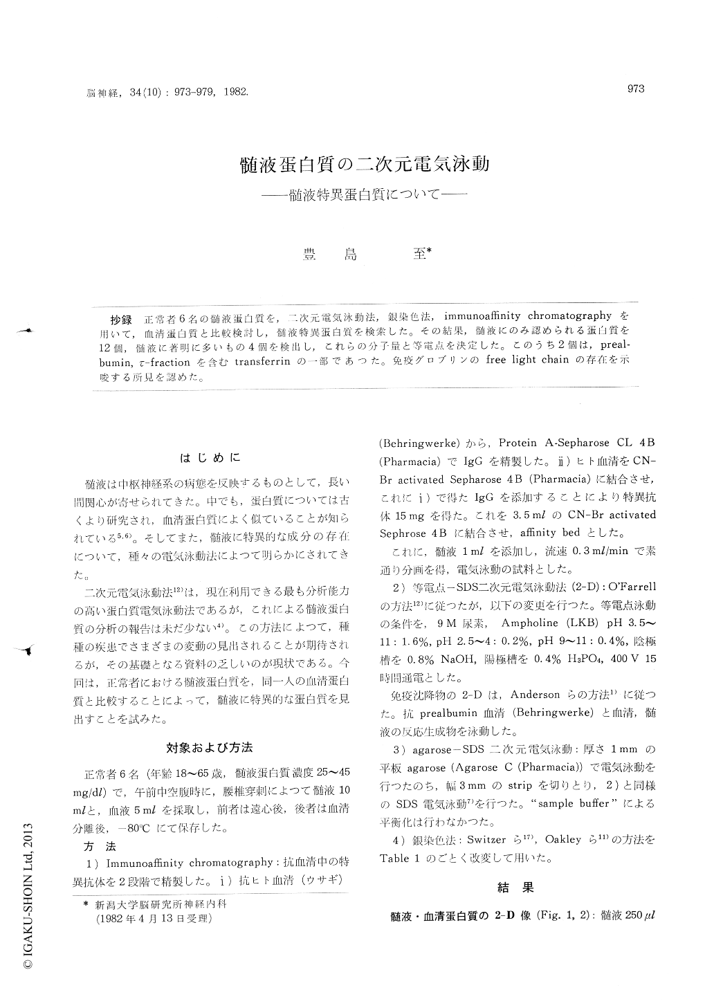
抄録 正常者6名の髄液蛋白質を,二次元電気泳動法,銀染色法,immunoaffinity chromatographyを用いて,血清蛋白質と比較検討し,髄液特異蛋白質を検索した。その結果,髄液にのみ認められる蛋白質を12個,髄液に著明に多いもの4個を検出し,これらの分子量と等電点を決定した。このうち2個は,preal—bumin,τ—fractionを含むtransferrinの一部であつた。免疫グロブリンのfree light chainの存在を示唆する所見を認めた。
Two-dimensional gel electrophoresis (2-D) and immunoaffinity chromatography (IAC) were employed to study the cerebrospinal fluid (CSF) characteristic proteins.
From the appearance of electrophoretic technique,CSF proteins have been analysed in relation to serum proteins. However, only recently the 2-D using silver staining was applied to CSF proteins (Goldman et al. (1980)). In the present study, the specificity of these proteins was further examined by IAC. This is the first report that combined the 2-D and IAC concerning the CSF proteins and determined the specificity of the CSF charac-teristic proteins accurately.
CSF proteins in six normal subjects were examined by 2-D. Isoelectric point (pI) and molecular weight (Mr) of these proteins were analysed by isoelectric focusing in the first dimension and SDS polyacrylamide gel electro-phoresis in the second dimension, respectively. Using this high resolution electrophoretic system and new silver staining technique, about 400 spots were resolved with high accuracy. Thus, a con-siderable increase (~30%) of the number of the resolved spots could be obtained with smaller (1/4) amount of proteins than those reported by Anderson and Anderson (1977). New procedure of silver staining is illustrated in Table 1.
2-D patterns obtained from 250 ,μl of CSF and 2.5μl of serum were presented in Fig. 1 (a) and (b), respectively. Corresponding diagrams are shown in Fig. 2. These patterns could be closely correlated with the 2-D map of plasma proteins published by Anderson and Anderson (1977).
Comparing these gel patterns, 16 characteristic spots were found on the 2-D map of CSF proteins ; 14 spots (A~L), which were not observed on the 2-D map of the serum, were clearly seen and 4 spots (a~d) were considerably increased in density on that of the CSF. Mr and pI of each spot wasestimated and listed in Table 2. Among these characteristic proteins, "b" (Mr: 17, 000, pI : 4. 8, 5.0, 5.3) and "J" (Mr: 74,000, 76,000, pI : 6. 2, 6. 3, 6.4) were identified as prealbumin and a species of transferrin, respectively. The latter is distinguished from that found in serum by the difference in pI and the most basic spot corres-ponds to so-called "r-fraction" Five of the six CSF characteristic proteins reported by Goldman et al. (1980) could be identified and are indicated in Table 2.
Further, immunoaffinity chromatography with anti-human serum antibodies was carried out. 1 ml of CSF was applied to an affinity column which bound 15 mg of specific antibodies against serum proteins. Employment of the specific antibodies rather than whole serum allowed us to reduce the bed volume considerably than that used by Wikkelso et al. (1978).
Unabsorbed fraction was collected and was subjected to 2-D analysis. About 60 spots were resolved on a silver stained gel, and, among them, 10 out of 16 CSF characteristic proteins and some of the serum components were distinguished. It is noteworthy that the light chains of immuno-globulins were observed while the heavy chains could not be detected. It suggests that free light chain exists in the CSF.
The method described here, combining immu-noaffinity chromatography and 2-D together with new silver staining method, will be readily applied to the investigation of the pathological CSF and pathological central and peripheral nervous tissue.

Copyright © 1982, Igaku-Shoin Ltd. All rights reserved.


