Japanese
English
- 有料閲覧
- Abstract 文献概要
- 1ページ目 Look Inside
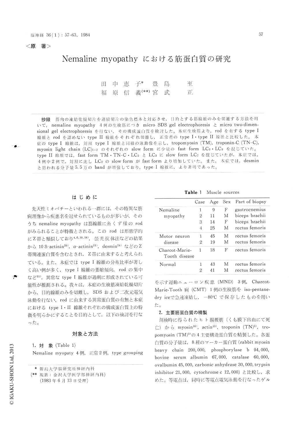
抄録 筋肉の凍結乾燥切片を連続切片の染色標本と対応させ,目的とする筋線維のみを切離する方法を用いて,nemaline myopathy 4例の生検筋につき micro SDS gel electrophoresisとmicro two-dimen—sional gel electrophoresisを行ない,その構成蛋白質を検討した。本症生検筋より, rodを有するtype I線維とrodを認めないtype II線維をそれぞれ切離し,正常者のtype I・type II線維と比較した。本症のtype I線維は,対照typeI線維と同様の泳動像を示し, tropomyosin (TM), troponin-C (TN-C),myosin light chain (LC)1・2のそれぞれのslow formに少量のfast form LC2・LC3を混じていた。type II線維では, fast form TM・TN-C・LC2とLC3にslow form LC2を混じていたが.本症では,4例中2例で,対照に比しLC2のslow formがfast formより増加していた。また,本症では,desminと思われる分子量5.5万のbandが増強しており, type I 線維に,より著明であった。
The characteristic features observed in the mus-cle fibers of nemaline myopathy are the presence of rods mainly in type I fibers, and the predomi-nance and atrophy of type I fibers.
In order to detect the abnormal proteins in the rods and clarify whether type I or II fibers have abnormal structural protein, we examined proteins in the muscles of patients with nemaline myopathy by one and two-dimensional gel electrophoresis (2-D). These freshly frozen muscles were cut to 20 μm thick and freeze-dried. Pieces were chosen and teased under a dissecting microscope with re-ference to the stained specimens of the same part, and electrophoresed.
At first, we examined the proteins of the type I and II fibers in normal and type grouping fibers of patients with motor neuron disease by SOS gel electrophoresis and found no abnormality of the protein pattern in this disease. Then we examined each of the following fibers of the nemaline mus-cle ; 1) type I fibers with many rods and 2) type II fibers with no rods. Each of the electrophoresed gel patterns of the nemaline muscle was compared with those of grouping type I and II fibers. The SDS gel electrophoresis showed and increase of the intensity of 55 K band in nemaline myopathy, especially in type I fibers with rods, which was thought as desmin. In 2-D, the pattern of type I fibers with rods were identical to that of the group-ing type I fibers except the 55 K spot which show-ed a slow form of tropomyosin α-subunit (TM-α), troponin-C (TN-C) and myosin light chains (LC) although both contained small amounts of fast form LC2 and LC3.
The 2-D pattern of type II fibers were compo-sed of fast form TM-α and myosin LC with a small amount of slow form LC2. This pattern was almost identical in nemaline muscle and control, but the intensity of the sLC2 spot was stronger than fLC2 in two of four patients with nemaline myopathy.
Our method of gel electrophoresis using freeze-dried tissue is superior in the following respect. The required fibers can be dissected with referen-ce to stained serial tissues at any time with the minimum risk of protein breakdown.

Copyright © 1984, Igaku-Shoin Ltd. All rights reserved.


