Japanese
English
- 有料閲覧
- Abstract 文献概要
- 1ページ目 Look Inside
I.アミロイド斑の形態学的側面
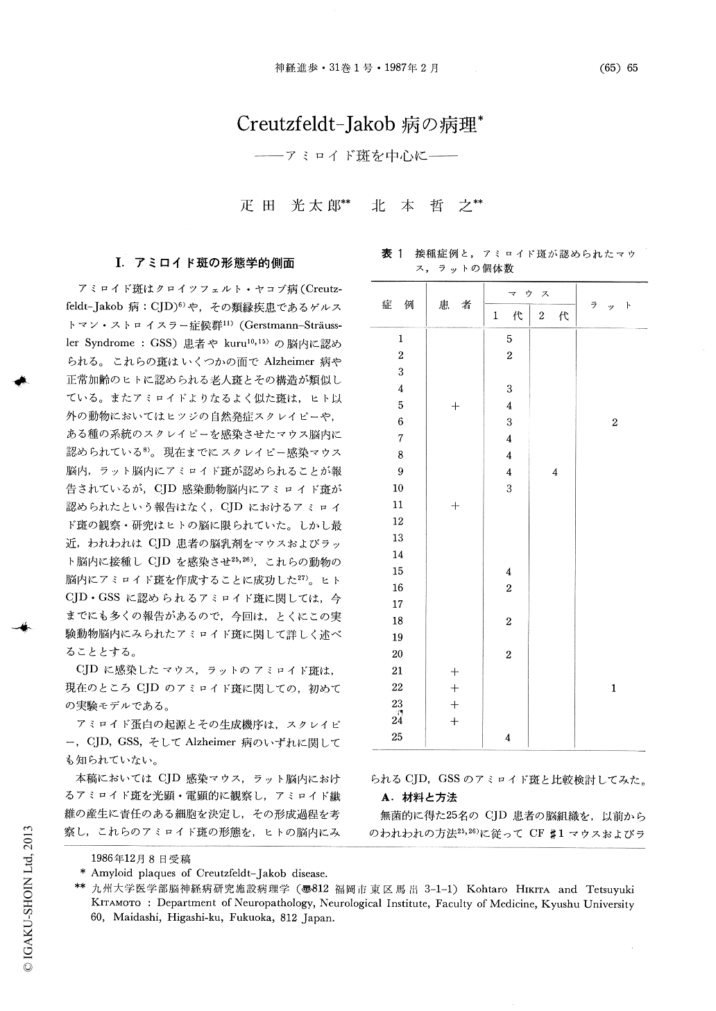
アミロイド斑はクロイツフェルト・ヤコブ病(Creutzfeldt-Jakob病:CJD)6)や,その類縁疾患であるゲルストマン・ストロイスラー症候群11)(Gerstmann-Sträussler Syndrome:GSS)患者やkuru10,15)の脳内に認められる。これらの斑はいくつかの面でAlzheimer病や正常加齢のヒトに認められる老人斑とその構造が類似している。またアミロイドよりなるよく似た斑は,ヒト以外の動物においてはヒツジの自然発症スクレイピーや,ある種の系統のスクレイピーを感染させたマウス脳内に認められている8)。現在までにスクレイピー感染マウス脳内,ラット脳内にアミロイド斑が認められることが報告されているが,CJD感染動物脳内にアミロイド斑が認められたという報告はなく,CJDにおけるアミロイド斑の観察・研究はヒトの脳に限られていた。しかし最近,われわれはCJD患者の脳乳剤をマウスおよびラット脳内に接種しCJDを感染させ25,26),これらの動物の脳内にアミロイド斑を作成することに成功した27)。ヒトCJD・GSSに認められるアミロイド斑に関しては,今までにも多くの報告があるので,今回は,とくにこの実験動物脳内にみられたアミロイド斑に関して詳しく述べることとする。
CJDに感染したマウス,ラットのアミロイド斑は,現在のところCJDのアミロイド斑に関しての,初めての実験モデルである。
1) Morphological aspects
Amyloid plaques were experimentally produced in the brains of mice inoculated with human Creutzfeldt-Jakob disease (CJD) brain homogenate. Light-microscopically, these plaques were mostly round and oval and were surrounded by macrophages and astrocytes. Ultramicroscopically, amyloid plaques were present in the cytoplasm of macrophages or were surrounded by these cells. The macrophages had numerous Golgi apparatus, endoplasmic reticula(ER), ribosomes, polysomesandlysosomes with inoculated materials or degenerating products. The bundles of amyloid fibrilswere intermingled with the cytoplasm of macro-absent. Some bundles of amyloid fibrils projected from the Golgi apparatus or rough ER and were partly exposed to the extracellular spaces, but there were no amyloid fibrils in the lysosomes. These findings confirmed that amyloid fibrils in the brains of CJD infected mice were produced by macrophages. Ultramicroscopical observation of human CJD and Gerstmann-Strdussler-Scheinker disease also revealed close relationship between amyloid plaques and macrophage.

Copyright © 1987, Igaku-Shoin Ltd. All rights reserved.


