Japanese
English
- 有料閲覧
PPV購入案内
1,320円 (1,200円+税10%) こちらは、481ページから487ページの文献です。購入には医書.jp本会員登録が必要です。
会員登録完了後に改めて上記「購入サイトへ」を選択してください。
または「購入サイトへ」選択後に「購入ログイン」、「新規会員登録」の順に進んでください。
会員登録について詳しくはをご確認ください。
- Abstract 文献概要
- 1ページ目 Look Inside
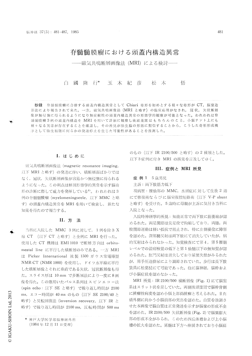
抄録 脊髄髄膜瘤に合併する頭蓋内構造異常としてChiari奇形を初めとする種々な奇形がCT,脳室造影法により報告されて来た。一方,磁気共鳴画像法(MRIと略す)の臨床応用がなされ,冠状,矢状断層像が無侵襲に得られるようになり無症候性の頭蓋内構造異常の形態学的観察が可能となった。われわれは脊髄髄膜瘤3例の頭蓋内構造をMRIを用いて詳細に観察し後頭蓋窩はもちろんのこと,小脳テント上にも種々なる異常が存在することを確認し,その所見が胎生脳の形態に類似することから,こうした奇形形成機序として胎生初期に何らかの発達停止を生じた可能性があることを推測した。
It is generally accepted that myelomeningocele frequently associates with A rnold-Chiari malfor-mation and other anomalies of the intracranial structures. The ventriculographic and CT findings of the patients with myelomeningocele has been reported. Magnetic resonance (MR) imaging is useful to observe the coronal and sagittal images of the brain in order to speculate the etiological mechanism of myelomeningocele and its associated anomalies.
We experienced three cases of myelomeningo-cele and reviewed their MR images using coronal and sagittal tomography in spin echo and inversion recovery technique. The morphological detail of MR images as to the intracranial structures waspresented.
Possible mechanism of the anomalous structures of the brain in myelomeningocele was also des-cribed.
Copyright © 1985, Igaku-Shoin Ltd. All rights reserved.