Japanese
English
- 有料閲覧
- Abstract 文献概要
- 1ページ目 Look Inside
I.はじめに
骨格筋細胞の神経筋接合部には,神経から放出されるアセチルコリン(ACh)を受容し,筋細胞膜のNa+とK+に対する透過性を変化させ,その結果筋細胞膜の興奮を起こすアセチルコリンレセプター(AChR)という蛋白が存在する。このような蛋白の存在することはかなり以前から示唆されていたが,その研究が近年著しく進んだのは,Bungarus multicinctusというヘビから得られた神経毒α-bungarotoxin(BTX)が,筋肉に対するAChの脱分極作用を阻害しAChRに特異的に結合することが明らかになって以来である。
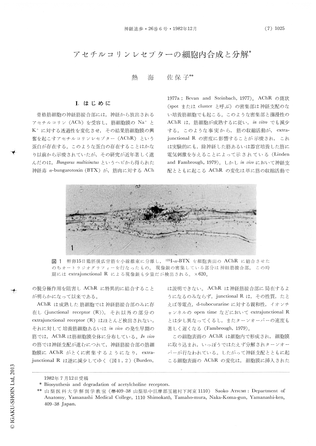
AChRは成熟した筋細胞では神経筋接合部のみに存在し(junctional receptor(R)),それ以外の部分のextrajunctional receptor(R)はほとんど検出されない。それに対して培養筋細胞あるいはin vivoの発生早期の筋では,AChRは筋細胞膜全体に分布している。In vivoの筋では神経支配が進むにつれて,神経筋接合部の筋細胞膜にAChRがとくに密集するようになり,extrajunctional Rは逆に減少してゆく(図1,2)(Burden,1977a;Bevan and Steinbach,1977)。AChRの斑状(spotまたはclusterと呼ぶ)の密集部は神経支配のない培養筋細胞でも起こる。
Intracellular synthesis and degradation of acetylcholine receptors (AChR) of skeletal muscle fibers were described by way of morphological studies which depended on 125I-α-bungarotoxin to detect the AChR. In vivo as well as in vitro, puromycin-sensitive silver grains for AChR were observed over the Golgi complex in latissimus dorsi muscles in 11-day incubated chick embryos. The Golgi complex is presumably involved in the glycosylation and assembly of AChR molecules. Multivesicular bodies also showed heavy grains covering them, which were sensitive to puromycin treatment.

Copyright © 1982, Igaku-Shoin Ltd. All rights reserved.


