Japanese
English
- 有料閲覧
- Abstract 文献概要
- 1ページ目 Look Inside
はじめに
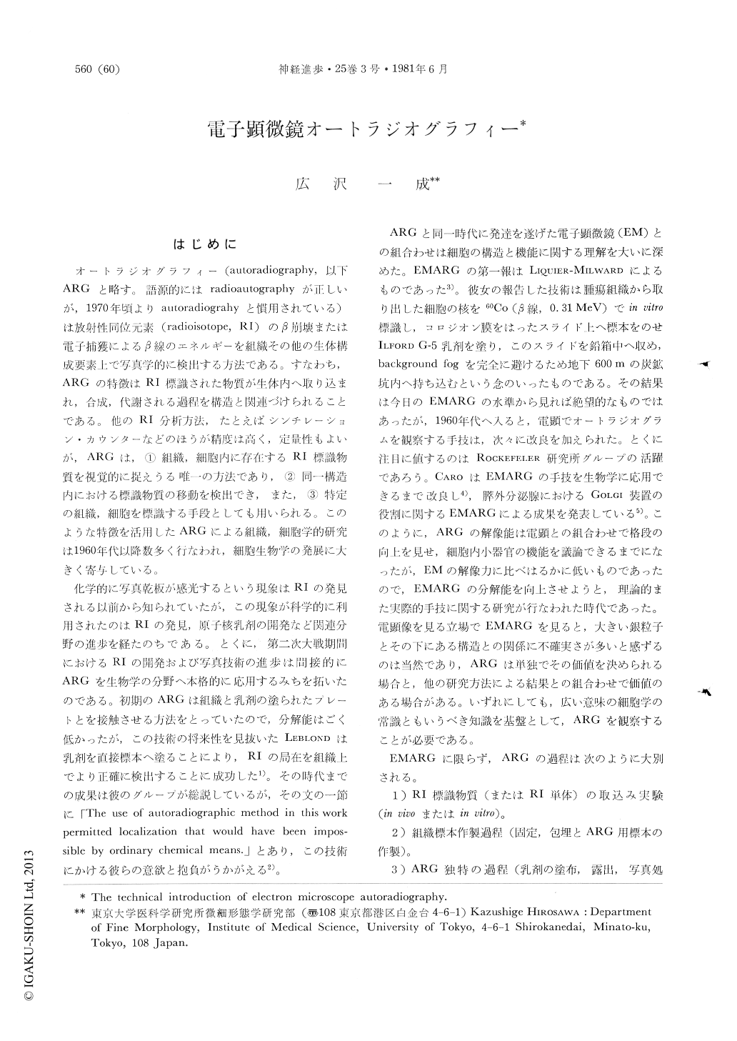
オートラジオグラフィー(autoradiography,以下ARGと略す。語源的にはradioautographyが正しいが,1970年頃よりautoradiograhyと慣用されている)は放射性同位元素(radioisotope,RI)のβ崩壊または電子捕獲によるβ線のエネルギーを組織その他の生体構成要素上で写真学的に検出する方法である。すなわち,ARGの特徴はRI標識された物質が生体内へ取り込まれ,合成,代謝される過程を構造と関連づけられることである。他のRI分析方法,たとえばシンチレーション・カウンターなどのほうが精度は高く,定量性もよいが,ARGは,①組織,細胞内に存在するRI標識物質を視覚的に捉えうる唯一の方法であり,②同一構造内における標識物質の移動を検出でき,また,③特定の組織,細胞を標識する手段としても用いられる。このような特徴を活用したARGによる組織,細胞学的研究は1960年代以降数多く行なわれ,細胞生物学の発展に大きく寄与している。
化学的に写真乾板が感光するという現象はRIの発見される以前から知られていたが,この現象が科学的に利用されたのはRIの発見,原子核乳剤の開発など関連分野の進歩を経たのちである。とくに,第二次大戦期間におけるRIの開発および写真技術の進歩は間接的にARGを生物学の分野へ本格的に応用するみちを拓いたのである。
Abstract
Electron microscope autoradiography was for the first time introduced by LIQUIER-MILWARD (1956). In early sixties, the authentic techniques were developed by CARO (1961) and others, which resulted in great participation of EMARG to modern cell biology. Among variations of EMARG techniques, a technique devised by SALPETER and BACHMANN (1964), "a dipping method", has bcen modified by many researchers including LARRA and DROTZ (1970), because the dipping method was most easy to manipulate. A modification of the method is introduced in this article with special reference to the precise technical procedures.

Copyright © 1981, Igaku-Shoin Ltd. All rights reserved.


