Japanese
English
- 有料閲覧
- Abstract 文献概要
- 1ページ目 Look Inside
I.はじめに
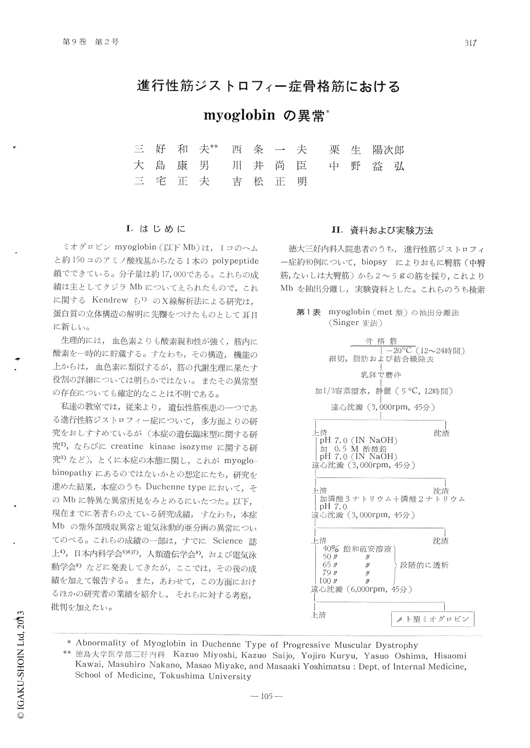
ミオグロビンmyoglobin(以下Mb)は,1コのヘムと約150コのアミノ酸残基からなる1本のpolypeptide鎖でできている。分子量は約17,000である。これらの成績は主としてクジラMbについてえられたもので,これに関するKendrewら1)のX線解析法による研究は,蛋白質の立体構造の解明に先鞭をつけたものとして耳目に新しい。
生理的には,血上色素よりも酸素親和性が強く,筋内に酸素を一時的に貯蔵する。すなわち,その構造,機能の上からは,血色素に類似するが,筋の代謝生理に果たす役割の詳細については明らかではない。またその異常型の存在についても確定的なことは不明である。
On the myoglobin (Mb) obtained by Singer'smethod from the biopsied muscle of the pa-tients with progressive muscular dystrophy(PMD), spectrophotometric examinations andelectrophoretic studies were made by thepresent authors.
Spectrophotometry in the visible regionrevealed no recognizable differences in theabsorption curves between the met-Mb fromthe patients with PMD and that from cotrols.However, in the ultraviolet region in Duche-nne type of PMD, the maximum of the abso-rption at pH 7.0 was approximately at 275mμin contrast to 281mμ in normal controls, neu-rogenic muscular atrophy and other types ofPMD.
The electrophoretic studies using celluloseacetate membrane and Cyanogum gel revealedthat the Mb of normal controls could be sepa-rated into four subfractions, Mb1, Mb2, Mb3, andMb4, being predominated by Mb1 among thefour. But, in Duchenne type of PMD, Mb1decreased markedly and Mb3 increased relati-vely. This abnormal electrophoretic patternof Mb subtraction was not recognized in Mbof normal and other controls. Absorption ma-xima in the ultraviolet region of Mb3 and Mb4usually shifted to the blue in contrast to thoseof Mb1 and Mb2 at 281mμ, and the characteristicchanges of Mb of Duchenne type of PMD areconsidered to be ascribed to the decrease ormalformation of Mb1 and a relative increaseof Mb3.
It is of interest to presume that the sub-fractions of Mb in the Duchenne type of PMDare so modified in amino acid composition orin three dimensional structures of protein ascompared with those of normal Mb.

Copyright © 1965, Igaku-Shoin Ltd. All rights reserved.


